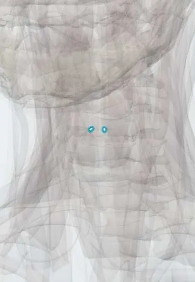
Skeletal system · Head and neck
Corniculate cartilages !
The corniculate cartilages are tiny horn-shaped cartilages on top of the arytenoids, supporting the folds at the back of the entrance to the larynx.
Official name: Corniculate cartilage
Opens in the Durgical app, no sign-up needed.
What it is
Tiny horn-shaped cartilages on top of the arytenoids.
What it does
Support the folds at the back of the larynx entrance.
Key facts
- Where
- On the apex of each arytenoid, in the aryepiglottic folds
- Function
- Support the back of the laryngeal inlet
Relations
- Below
- Apex of each arytenoid cartilage, to which they are joined or fused
- Lateral
- Cuneiform cartilages, further forward in the aryepiglottic fold
- Medial
- Interarytenoid notch at the back of the laryngeal inlet
- Behind
- Laryngopharynx; the pyriform recesses lie lateral to the aryepiglottic folds
How to find it
Not palpable. On laryngoscopy they are seen on the posterior rim of the laryngeal inlet as two small rounded tubercles on top of the arytenoid mounds.
On imaging
Too small to judge on their own; laryngoscopy shows them as the corniculate tubercles at the back of the laryngeal inlet. On contrast CT or MRI of the neck they are small bumps on the tips of the arytenoids, at the back of the aryepiglottic folds. Imaging assesses this area rather than the cartilages: tumour spread along the aryepiglottic folds or swelling of the back of the larynx.
Clinical notes
- Airway landmark: in a poor laryngoscopic view (Cormack–Lehane grade 2b) the corniculate and arytenoid tubercles may be all that shows behind the epiglottis
- In laryngomalacia, short aryepiglottic folds and loose mucosa over the arytenoid and corniculate region fall into the airway on breathing in, causing stridor in infants
- Angioedema or anaphylaxis swells the loose mucosa over the corniculate and cuneiform tubercles and along the aryepiglottic folds, narrowing the laryngeal inlet and causing stridor
Test yourself
Think of the answer, then tap the question to see it.
Where are the corniculate cartilages and what do they support?
On the apex of each arytenoid cartilage, in the aryepiglottic folds; with the cuneiform cartilages they stiffen the back of the aryepiglottic folds and the laryngeal inlet.
Why do the corniculate cartilages not ossify with age, unlike the thyroid and cricoid cartilages?
They are elastic cartilage, like the epiglottis; the thyroid and cricoid are hyaline cartilage, which ossifies.
In a poor laryngoscopic view (Cormack–Lehane grade 2b), what may be all that shows behind the epiglottis?
The corniculate and arytenoid tubercles at the back of the laryngeal inlet.
Educational information, not medical advice. For any symptom, see a health professional.
Its names
- Official Latin (TA2)
- cartilago corniculata TA2 997
- Official Spanish (DeCS)
- Cartílagos corniculados
- English (TA2)
- Corniculate cartilage
- Also called
- Santorini's cartilage · cartilago Santorini
Sources: Terminologia Anatomica (TA2); Anatomy, Head and Neck: Larynx Cartilage; Anatomy, Head and Neck: Larynx.
Spotted a mistake?We are always improving for the community. Tell us what is wrong and our team will review it.Report a mistake
Representative model, for educational purposes only.