Respiratory system · Head and neck
Epiglottis !
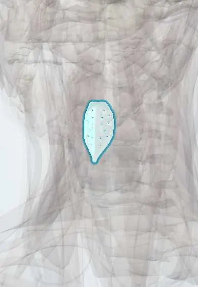
The epiglottis is a leaf-shaped flap of cartilage behind the tongue; it tips back over the larynx when swallowing so food goes down the oesophagus.
Opens in the Durgical app, no sign-up needed.
What it is
A leaf-shaped flap of elastic cartilage behind the tongue.
What it does
Tips back over the larynx when swallowing so food goes to the oesophagus.
Key facts
- Where
- Behind the tongue base, top of the larynx
- Blood supply
- Superior laryngeal artery
- Nerve
- Internal laryngeal nerve (X)
- Function
- Covers the laryngeal inlet when swallowing
Relations
- In front
- Valleculae, the paired pockets between it and the tongue base, either side of the median glossoepiglottic fold; lower down the pre-epiglottic space, with fat and the hyoepiglottic ligament
- Below
- Petiole, attached by the thyroepiglottic ligament to the inner angle of the thyroid cartilage just above the vocal folds
- Lateral
- Aryepiglottic folds running back to the arytenoid cartilages; the quadrangular membrane hangs from its sides
- Behind
- Laryngeal vestibule and, below it, the vocal folds
How to find it
A laryngeal mirror or flexible endoscope shows its free edge behind the tongue base, with the valleculae in front of it, bounded by the median and lateral glossoepiglottic folds; during intubation it is the first landmark the laryngoscope blade looks for.
On imaging
Lateral neck radiograph in epiglottitis: a swollen, rounded epiglottis (thumb sign), thick aryepiglottic folds and loss of the air pocket in the vallecula (vallecula sign). Imaging is not allowed to delay securing the airway.
Common problems
- Epiglottitis: rare but dangerous swelling that can block the airway (drooling, muffled voice).
- When it does not close well, food or liquid goes into the airway (aspiration).
How it is diagnosed
Careful airway exam by a specialist; swallowing studies.
Treatment
- Securing the airway and antibiotics for epiglottitis.
- Swallowing therapy.
Clinical notes
- Laryngoscopy: a curved blade tip goes into the vallecula and lifts the epiglottis indirectly; a straight blade lifts it directly, often used in infants with a long, floppy epiglottis
- The pre-epiglottic space in front of it is a route of spread for supraglottic cancer and changes staging and surgery
- A curled, omega-shaped epiglottis in infants is part of laryngomalacia, the most common cause of inspiratory stridor in infancy
Development
Arises from the hypobranchial eminence, behind the developing tongue, at the level of the fourth pharyngeal arch.
Related conditions
- Laryngeal cancer
- Anaphylaxis
- Foreign-body airway obstruction (choking)
- Hymenoptera stings (bees, wasps, ants)
- Oropharyngeal dysphagia in older adults
- Acute epiglottitis
Test yourself
Think of the answer, then tap the question to see it.
What is the classic presentation of acute epiglottitis in a child?
Fever, sore throat, drooling, muffled voice and sitting forward in the tripod position; the airway is secured in a controlled setting before the throat is examined.
What is the thumb sign on a lateral neck radiograph?
A swollen, rounded epiglottis in epiglottitis, often with thick aryepiglottic folds and loss of the vallecular air pocket; imaging must not delay securing the airway.
How do curved and straight laryngoscope blades lift the epiglottis?
A curved blade tip goes into the vallecula and lifts it indirectly; a straight blade lifts the epiglottis directly, often used in infants with a long, floppy epiglottis.
Which vaccine made acute epiglottitis much less common in children?
Haemophilus influenzae type b (Hib) vaccine; adults now make up a larger share of cases.
What nerve gives sensation to the epiglottis and what space in front of it matters in supraglottic cancer?
The internal laryngeal nerve (X); the pre-epiglottic space in front of it is a route of tumour spread that changes staging and surgery.
Educational information, not medical advice. For any symptom, see a health professional.
Its names
- Official Latin (TA2)
- epiglottis TA2 3190
- Official Spanish (DeCS)
- Epiglotis DeCS D004825
- English (TA2)
- Epiglottis
Related
Sources: Terminologia Anatomica (TA2); DeCS; Epiglottitis; Anatomy, Head and Neck: Larynx Cartilage; Anatomy of the Human Body (20th ed.): Surface Markings of Special Regions of the Head and Neck (Lea & Febiger (Gray's Anatomy, 1918), 1918) · adapted, Public domain.
Spotted a mistake?We are always improving for the community. Tell us what is wrong and our team will review it.Report a mistake
Representative model, for educational purposes only.