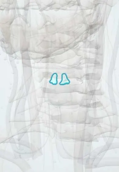
Skeletal system · Head and neck
Arytenoid cartilages !
The arytenoid cartilages are two small pyramid-shaped cartilages at the back of the larynx (voice box); they swivel to open and close the vocal folds.
Official name: Arytenoid cartilage
Opens in the Durgical app, no sign-up needed.
What it is
Two small pyramid cartilages on the back of the cricoid.
What it does
Swivel to open and close the vocal folds.
Key facts
- Where
- Paired pyramids on top of the cricoid lamina
- Function
- Vocal process anchors the vocal fold, muscular process takes the cricoarytenoid muscles; they rotate and slide to open and close the glottis
Relations
- Below
- Sloping upper border of the cricoid lamina, at the synovial cricoarytenoid joint
- Above
- Corniculate cartilages on each apex, inside the aryepiglottic folds
- In front
- Vocal process, from which the vocal ligament and the vocalis run forward to the thyroid cartilage; the vestibular ligament attaches just above it
- Medial
- The opposite arytenoid, across the interarytenoid region at the back of the glottis
- Behind
- Transverse and oblique arytenoid muscles, covered by the mucosa of the laryngopharynx
- Lateral
- Muscular process: the posterior cricoarytenoid pulls it from behind, the lateral cricoarytenoid from the front
How to find it
Not palpable. On laryngoscopy they are the two mounds at the back of the glottis, capped by the corniculate tubercles; how they move on breathing in and on a sustained vowel is how vocal fold mobility is judged.
On imaging
CT shows them at the level of the glottis. On the side of a vocal fold paralysis the arytenoid tends to tip forward and inward, with a wider pyriform sinus and laryngeal ventricle on that side.
Clinical notes
- Contact ulcer or intubation granuloma forms on the mucosa over the vocal process, where a tracheal tube or forceful voice use rubs
- Laryngeal electromyography helps tell a fixed joint (dislocation, arthritis) from a nerve palsy when a vocal fold does not move
- In bilateral vocal fold paralysis, removing part of one arytenoid (arytenoidectomy) widens the posterior glottis for breathing
- Cricoarytenoid joint arthritis, as in rheumatoid arthritis, can stiffen the joint and cause hoarseness or stridor
Development
They arise from sixth-arch cartilage together with the cricoid; the body is hyaline cartilage that may ossify with age, while the tip of the vocal process is elastic cartilage.
Related conditions
Test yourself
Think of the answer, then tap the question to see it.
What attaches to the vocal process and the muscular process of the arytenoid cartilages?
Vocal process: the back end of the vocal ligament. Muscular process: the posterior and lateral cricoarytenoid muscles.
What can arytenoid cartilage dislocation after intubation be mistaken for?
Recurrent laryngeal nerve palsy; both give a hoarse, breathy voice.
Which test helps tell a fixed arytenoid joint from a nerve palsy when a vocal fold does not move?
Laryngeal electromyography.
Where does an intubation granuloma form over the arytenoid cartilages?
On the mucosa over the vocal process, where a tracheal tube or forceful voice use rubs.
Educational information, not medical advice. For any symptom, see a health professional.
Its names
- Official Latin (TA2)
- cartilago arytenoidea TA2 983
- Official Spanish (DeCS)
- Cartílago Aritenoides DeCS D001193
- English (TA2)
- Arytenoid cartilage
- Also called
- cartilago arytaenoidea · cartilago arytaenoides
Related
Sources: Terminologia Anatomica (TA2); DeCS; Anatomy, Head and Neck: Larynx Arytenoid Cartilage; Anatomy, Head and Neck: Larynx Cartilage; Anatomy of the Human Body (20th ed.): The Branchial Region (Lea & Febiger (Gray's Anatomy, 1918), 1918) · adapted, Public domain.
Spotted a mistake?We are always improving for the community. Tell us what is wrong and our team will review it.Report a mistake
Representative model, for educational purposes only.