Digestive system · Head and neck
Lower right central incisor !
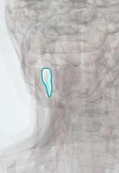
The lower right central incisor (tooth 41) is a small flat front tooth beside the midline that cuts food.
Opens in the Durgical app, no sign-up needed.
What it is
A front tooth with a thin, straight cutting edge and one root.
What it does
Bites into and cuts food; shapes some speech sounds (f, v, th).
Key facts
- Joints with
- Alveolar socket of the mandible (gomphosis held by the periodontal ligament); bites against the opposing teeth
- Landmarks
- FDI 41, lower right central incisor: 1 root; chisel-shaped crown for cutting; erupts at about 6–7 years; nerve: inferior alveolar nerve (V3)
Relations
- Medial
- The lower left central incisor
- Lateral
- The lower right lateral incisor
- Above
- Meets only the upper right central incisor, which overlaps it in front
- In front
- Lower lip, the mentalis and a thin plate of labial bone
How to find it
First tooth right of the lower midline (the patient's right); it sits behind the upper centrals in a normal bite, so the lower lip must be pulled down to see its face.
On imaging
Periapical films are the study of choice; a second canal shows only when the beam is angled from the side. Bone loss between the lower incisors is easy to follow on serial periapicals.
Common problems
- Chipped or broken front teeth after falls or blows.
Clinical notes
- Incisive branch of the inferior alveolar nerve (V3); nerve fibres from the opposite side cross the midline, and thin labial bone lets infiltration numb it
- Lower incisors commonly crowd during adolescence and adult life, even after orthodontic treatment
- With the first molars, it is a typical site of the molar-incisor pattern of periodontitis seen in young people
Development
Replaces the primary lower right central incisor (FDI 81); with the first molars, the lower central incisors usually open the mixed dentition.
Related conditions
Test yourself
Think of the answer, then tap the question to see it.
Where does tartar build up fastest on the lower right central incisor, and why?
On its lingual surface, opposite the openings of the submandibular ducts under the tongue.
Why can a second canal be missed in the lower right central incisor?
Its root is flattened side to side and often holds 2 canals (labial and lingual), easy to miss on a front-on radiograph.
Which nerve supplies the lower right central incisor?
The incisive branch of the inferior alveolar nerve (V3), with fibres crossing from the opposite side at the midline.
Which teeth are involved in the molar-incisor pattern of periodontitis in young people?
The incisors, such as the lower right central incisor, and the first molars.
Educational information, not medical advice. For any symptom, see a health professional.
Related
Sources: Anatomy, Head and Neck, Teeth; Anatomy, Permanent Dentition; Anatomy, Head and Neck, Tooth Eruption; Anatomy, Head and Neck: Alveolar Nerve; 23.3 The Mouth, Pharynx, and Esophagus (OpenStax (Rice University), 2022) · adapted, CC BY 4.0.
Spotted a mistake?We are always improving for the community. Tell us what is wrong and our team will review it.Report a mistake
Representative model, for educational purposes only.