Cardiovascular system · Upper limb
Median cubital vein !
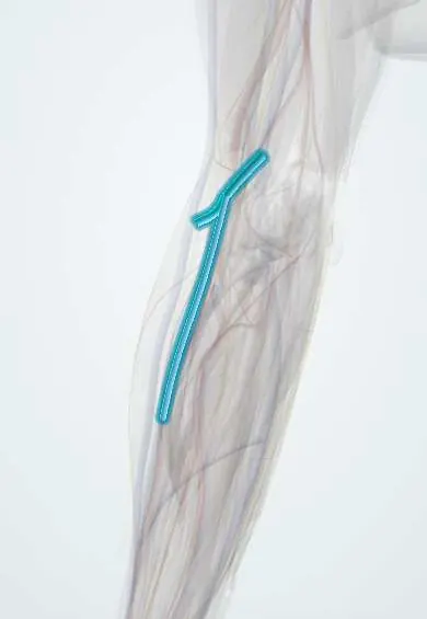
The median cubital vein crosses the front of the elbow, linking the cephalic and basilic veins; it is the usual vein for blood draws.
Opens in the Durgical app, no sign-up needed.
What it is
The surface vein that crosses the front of the elbow between the cephalic and basilic veins.
What it does
The usual vein for blood draws.
Key facts
- Drains
- Cephalic vein at the elbow
- Into
- Basilic vein (crosses the cubital fossa over the bicipital aponeurosis)
Relations
- Course
- Leaves the cephalic vein a little below the elbow crease and runs up and medially across the cubital fossa to the basilic vein above the medial epicondyle
- Deep
- Bicipital aponeurosis; beneath it the brachial artery and the median nerve
- Superficial
- Skin, with branches of the medial and lateral antebrachial cutaneous nerves in the fat around it
How to find it
With the elbow straight and a tourniquet on the arm, it is seen or felt as a soft, springy tube crossing the elbow crease; ultrasound helps when it cannot be seen.
On imaging
Usually seen and felt without imaging. Ultrasound helps in obese patients or difficult venepuncture, showing the vein over the bicipital aponeurosis, which separates it from the brachial artery and median nerve lying deeper. The vein compresses fully under the probe and the artery does not. After a misplaced puncture, ultrasound shows a haematoma or a false aneurysm of the artery.
Common problems
- The usual site for blood draws and IV lines at the elbow crease.
- Bruising or phlebitis after needle sticks; rarely a nerve or the brachial artery is irritated beneath it.
How it is diagnosed
Seen and felt after placing a tourniquet; ultrasound helps when veins are hard to find.
Treatment
- Warm compresses and arm elevation for bruising or mild phlebitis.
Clinical notes
- A deep or very medial needle can pass through the aponeurosis and reach the brachial artery or the median nerve
- A superficial or high-origin artery at the elbow can lie where a vein is expected; a pulsating vessel is not a vein to puncture
- Common site for transfusions, contrast injections and short IV lines when the forearm veins are poor
Anatomical variants
- Patterns vary: the classic oblique link from cephalic to basilic, an M-shaped pattern in which the median antebrachial vein splits into median cephalic and median basilic veins, or no median cubital vein at all; studies sort them into several types
Test yourself
Think of the answer, then tap the question to see it.
Why is the median cubital vein the safest common vein for blood draws?
The bicipital aponeurosis separates it from the brachial artery and median nerve.
Why does the median cubital vein roll less under the needle?
A perforating vein links it to the deep veins and anchors it.
What usually causes a shooting pain during a draw from the median cubital vein?
Branches of the medial and lateral antebrachial cutaneous nerves passing close to it.
Educational information, not medical advice. For any symptom, see a health professional.
Its names
- Official Latin (TA2)
- vena mediana cubiti TA2 4980
- Official Spanish (DeCS)
- Vena mediana del codo
- English (TA2)
- Median cubital vein
Related
Sources: Terminologia Anatomica (TA2); Anatomy, Shoulder and Upper Limb, Elbow Cubital Fossa; Anatomy, Shoulder and Upper Limb, Veins.
Spotted a mistake?We are always improving for the community. Tell us what is wrong and our team will review it.Report a mistake
Representative model, for educational purposes only.