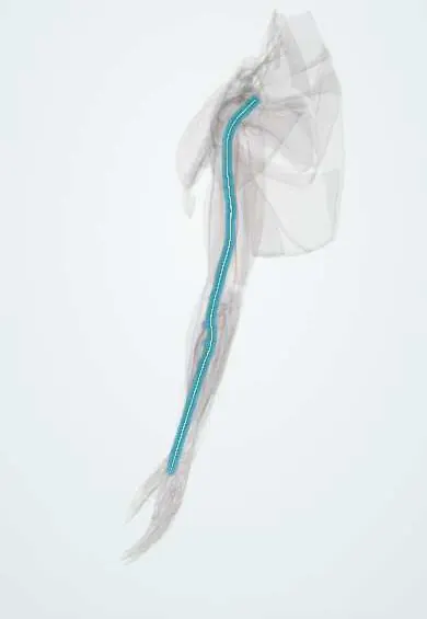
Median nerve !
The median nerve runs down the middle of the arm and through the carpal tunnel; it gives feeling to the thumb side of the hand and moves the thumb pad.
Opens in the Durgical app, no sign-up needed.
What it is
The nerve down the middle of the arm and forearm that passes through the carpal tunnel.
What it does
Feeling in the thumb, index, middle and half the ring finger; powers most forearm flexors and the thumb pad.
Key facts
- Roots
- C5(6)–T1, from the lateral and medial cords
- Moves
- Pronator teres, FCR, palmaris longus, FDS; via AIN: FPL, FDP (index, middle), pronator quadratus; thenar muscles (APB, OP, superficial FPB); lumbricals 1–2
- Feeling from
- Palmar thumb, index, middle and lateral half of the ring finger (and their dorsal tips); lateral palm
Relations
- Course
- Formed by its two roots in front of the axillary artery, it runs lateral to the brachial artery, crosses it at mid-arm and lies medial to it at the elbow; it passes between the heads of pronator teres, runs between FDS and FDP, becomes superficial just above the wrist and enters the carpal tunnel
- In front
- At the elbow, the bicipital aponeurosis and the median cubital vein; at the wrist, the palmaris longus tendon (when present) and the flexor retinaculum
- Behind
- Brachialis at the elbow; flexor digitorum profundus in the forearm
- Lateral
- The biceps tendon and brachial artery in the cubital fossa; the flexor carpi radialis tendon at the wrist
- Medial
- The flexor digitorum superficialis tendons at the wrist
How to find it
At the elbow it lies just medial to the brachial artery pulse, which is itself medial to the biceps tendon (made prominent by resisted elbow flexion). At the wrist it lies under or just radial to the palmaris longus tendon, between it and the flexor carpi radialis tendon.
Examination
- Thumb palmar abduction against resistance (abductor pollicis brevis, 'pen test'): the most useful motor test of the median nerve at the wrist
- Phalen test: wrists held fully flexed for about a minute bring on tingling in the median fingers, supporting carpal tunnel syndrome; Tinel's sign and the carpal compression (Durkan) test point the same way
- Pinprick or two-point discrimination at the tip of the index finger, the area supplied by the median nerve alone
- Pronator compression test: firm pressure over the proximal pronator teres reproducing tingling, with palm numbness, suggests pronator syndrome rather than carpal tunnel syndrome
On imaging
Ultrasound is the usual first image. In carpal tunnel syndrome the nerve is swollen and hypoechoic at the tunnel inlet (pisiform level), where its cross-sectional area is measured, and flat under the retinaculum. Ultrasound or MRI also shows masses, a bifid nerve or a persistent median artery. Nerve conduction studies remain the reference; MRI shows thenar denervation.
Common problems
- Carpal tunnel syndrome: squeezed at the wrist, the most common nerve compression in the body.
- Injured by cuts at the wrist or by fractures near the elbow in children.
- Weak, wasted thumb pad in long-standing compression.
How it is diagnosed
Feeling in the thumb, index and middle fingers, thumb strength, Tinel and Phalen tests. Nerve conduction studies and EMG.
Treatment
- Splint, activity changes and injection for mild carpal tunnel syndrome.
- Carpal tunnel release when symptoms are constant or the nerve is damaged.
- Microsurgical repair after a cut.
Clinical notes
- Extension-type supracondylar humerus fracture in children: posterolateral displacement of the distal fragment stretches the median nerve (most often its AIN fibres) and the brachial artery over the proximal fragment
- Pronator syndrome: compression at the elbow (ligament of Struthers, bicipital aponeurosis, pronator teres, FDS arch) gives forearm ache and palm-base numbness, which carpal tunnel syndrome spares
- Carpal tunnel release divides the flexor retinaculum on the ulnar side of the nerve, away from the recurrent motor branch and the palmar cutaneous branch
- Wrist lacerations from glass or knives cut it between the palmaris longus and flexor carpi radialis tendons, often with flexor tendons
- Deep venipuncture on the medial side of the cubital fossa can reach it, although the bicipital aponeurosis usually shields it
Anatomical variants
- Martin-Gruber anastomosis: a branch from the median or anterior interosseous nerve to the ulnar nerve in the forearm, so some 'ulnar' hand muscles keep working after a high ulnar lesion
- Supracondylar process of the humerus with a ligament of Struthers to the medial epicondyle; the nerve, sometimes with the brachial artery, passes under it and can be compressed
- Bifid median nerve in the carpal tunnel, often with a persistent median artery between the two halves
- Riche-Cannieu anastomosis in the palm, between the recurrent motor branch and the deep branch of the ulnar nerve
Development
In the embryo a median artery runs with the nerve and is the main artery of the forearm; it normally regresses as the radial and ulnar arteries enlarge. When it persists it follows the nerve into the palm, sometimes between the halves of a bifid nerve, and can add to carpal tunnel compression.
Memory aid
Cubital fossa, lateral to medial: 'TAN' (Tendon of biceps, Artery, Nerve). Median muscles in the hand: 'LOAF' (Lumbricals 1–2, Opponens pollicis, Abductor pollicis brevis, Flexor pollicis brevis).
Related conditions
Test yourself
Think of the answer, then tap the question to see it.
In the cubital fossa, where does the median nerve lie relative to the biceps tendon and brachial artery?
Most medial: biceps tendon, then brachial artery, then median nerve, lateral to medial.
How does the hand look after a median nerve lesion at the elbow versus at the wrist?
At or above the elbow, «hand of benediction» on making a fist (index and middle stay straight); at the wrist, thenar wasting with the thumb in line with the fingers («ape hand»).
What is the most useful motor test of the median nerve at the wrist?
Resisted palmar abduction of the thumb (abductor pollicis brevis, «pen test»).
What symptom separates pronator syndrome from carpal tunnel syndrome in median nerve compression?
Numbness of the palm base: the palmar cutaneous branch leaves before the carpal tunnel, so carpal tunnel syndrome spares it while pronator syndrome does not.
Which childhood fracture classically stretches the median nerve, and which part?
The extension-type supracondylar humerus fracture with posterolateral displacement; most often its anterior interosseous fibres, with the brachial artery.
Educational information, not medical advice. For any symptom, see a health professional.
Its names
- Official Latin (TA2)
- nervus medianus TA2 6459
- Official Spanish (DeCS)
- Nervio mediano
- English (TA2)
- Median nerve
Related
- Brachial artery
- Flexor retinaculum
- FDS tendons
- FPL tendon
- Carpal tunnel
- Cubital fossa
- Pronator teres
- Flexor carpi radialis
Sources: Terminologia Anatomica (TA2); Anatomy, Shoulder and Upper Limb, Median Nerve; Anatomy, Shoulder and Upper Limb, Wrist Flexor Retinaculum; Anatomy of the Human Body (20th ed.): Development of the Vascular System (Lea & Febiger (Gray's Anatomy, 1918), 1918) · adapted, Public domain; Anatomía humana (4.ª ed.), tomo 1, cap. 62: Nervios del miembro superior.
Spotted a mistake?We are always improving for the community. Tell us what is wrong and our team will review it.Report a mistake
Representative model, for educational purposes only.