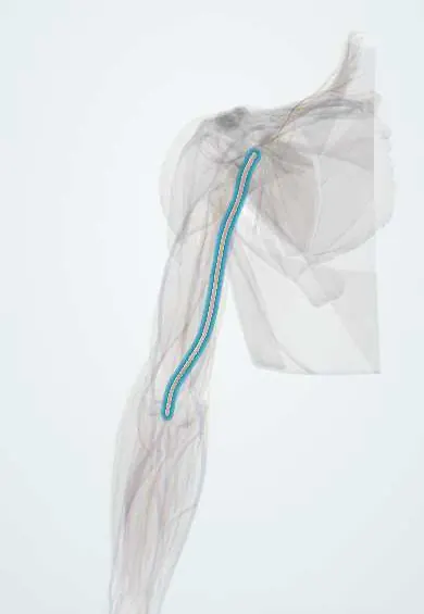
Cardiovascular system · Upper limb
Brachial artery !
The brachial artery is the main artery of the upper arm; blood pressure is measured over it and its pulse is felt at the inner elbow.
Opens in the Durgical app, no sign-up needed.
What it is
The main artery of the upper arm, along the inner side of the biceps to the elbow crease.
What it does
Feeds the arm; it is where blood pressure is measured and the elbow pulse is felt.
Key facts
- Origin
- Continuation of the axillary artery at teres major
- Branches
- Deep brachial, superior and inferior ulnar collateral, nutrient; ends as radial and ulnar arteries in the cubital fossa
- Supplies
- Arm (anterior compartment) and the elbow anastomosis
Relations
- Course
- From the lower border of teres major it descends along the medial side of the arm in the groove between the biceps and the coracobrachialis, then the brachialis, moving gradually in front of the humerus into the cubital fossa
- In front
- Skin and fascia for most of its length; the median nerve crossing at mid-arm; the bicipital aponeurosis and the median cubital vein at the elbow
- Behind
- Long and medial heads of the triceps, the insertion of coracobrachialis, then the brachialis; the radial nerve and deep brachial artery leave it backward at the top
- Medial
- Ulnar nerve in the upper half of the arm, until it pierces the medial intermuscular septum; basilic vein; median nerve at the elbow
- Lateral
- Coracobrachialis and biceps; median nerve in the upper arm; the biceps tendon in the cubital fossa
How to find it
Brachial pulse: in the cubital fossa just medial to the biceps tendon, with the elbow straight; higher up, in the groove along the medial border of the biceps. For blood pressure the cuff compresses it and the stethoscope is placed over it at the elbow. Its course is marked on the skin by a line from the apex of the axilla to the middle of the cubital fossa.
On imaging
Doppler ultrasound shows it at the elbow just medial to the biceps tendon, with the median nerve on its medial side; this is also a site for arterial access. In a child's displaced supracondylar fracture the radiograph shows the fracture, and a pulseless hand is assessed clinically and with Doppler; angiography is kept for selected cases. CT angiography is used in arm trauma.
Common problems
- Injured in elbow fractures (especially supracondylar fractures in children) and dislocations.
- Used to measure blood pressure and as an access point for some catheter procedures.
How it is diagnosed
Pulse and colour checks of the hand; Doppler ultrasound; CT angiography if an injury is suspected.
Treatment
- Urgent fracture reduction, which often restores the pulse.
- Vascular repair if the artery is injured.
Clinical notes
- Supracondylar humerus fracture in children: in the extension type the proximal fragment can kink or tear the artery and stretch the median nerve; a pale, poorly perfused hand is a surgical emergency
- Missed ischemia or compartment syndrome after these injuries can end in Volkmann ischemic contracture of the forearm flexors
- Ligation below the origin of the deep brachial artery is usually tolerated thanks to the elbow anastomosis; above it the risk of ischemia is higher
- A deep needle during venipuncture at the elbow can pass through the bicipital aponeurosis and reach the artery
Anatomical variants
- High division: it may split into the radial and ulnar arteries well above the elbow, in roughly 1 arm in 10 (reports vary), most often in the upper third of the arm and sometimes as high as the axilla; usually on one side only
- Superficial brachial artery: a vessel running in front of the median nerve instead of behind it, often continuing as a high-origin radial artery
- Supracondylar process: a small bony spur above the medial epicondyle, sometimes joined to it by a band (ligament of Struthers) under which the median nerve and brachial artery pass; a rare cause of compression
Development
It is part of the axis artery of the limb, which continues into the forearm as the anterior interosseous artery. The radial and ulnar arteries bud from it later, so a division above the elbow (high division) is a common variant.
Memory aid
Cubital fossa from lateral to medial (English): "Really Need Booze To Be At My Nicest": Radial Nerve, Biceps Tendon, Brachial Artery, Median Nerve.
Related conditions
Test yourself
Think of the answer, then tap the question to see it.
How does the median nerve relate to the brachial artery along the arm?
Lateral to it in the upper arm, crossing in front of it (usually) at mid-arm, and medial to it at the elbow.
What separates the brachial artery from the median cubital vein in the cubital fossa?
The bicipital aponeurosis; the artery lies medial to the biceps tendon.
Where does the brachial artery usually divide?
Into the radial and ulnar arteries at about the level of the neck of the radius.
Which childhood fracture threatens the brachial artery, and what late complication can follow missed ischemia?
The extension-type supracondylar humerus fracture; missed ischemia or compartment syndrome can end in Volkmann ischemic contracture.
Why is brachial artery ligation below the deep brachial artery usually tolerated?
The elbow anastomosis supplies the forearm; above that origin the risk of ischemia is higher.
Educational information, not medical advice. For any symptom, see a health professional.
Its names
- Official Latin (TA2)
- arteria brachialis TA2 4632
- Official Spanish (DeCS)
- Arteria braquial
- English (TA2)
- Brachial artery
Related
- Median nerve
- Brachial veins
- Brachial nodes
- Cubital fossa
- Biceps (long head)
- Biceps (short head)
- Brachialis
- Coracobrachialis
Sources: Terminologia Anatomica (TA2); Anatomy, Shoulder and Upper Limb, Brachial Artery; Anatomy of the Human Body (20th ed.): Development of the Vascular System (Lea & Febiger (Gray's Anatomy, 1918), 1918) · adapted, Public domain; Anatomía humana (4.ª ed.), tomo 1, cap. 59: Arterias del miembro superior; Tratado de anatomía humana (9.ª ed.), tomo II, Libro IV Angiología, arterias subclavia, axilar, humeral, radial y cubital, arcos palmares y vías anastomóticas del miembro superior.
Spotted a mistake?We are always improving for the community. Tell us what is wrong and our team will review it.Report a mistake
Representative model, for educational purposes only.