Reproductive system · Pelvis · Female
Mesosalpinx !
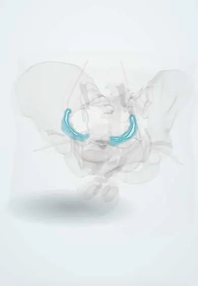
The mesosalpinx is the upper part of the broad ligament that holds the uterine tube and carries its blood vessels.
Opens in the Durgical app, no sign-up needed.
What it is
The part of the broad ligament that holds the tube.
What it does
Carries the tube's vessels; it is divided in a salpingectomy.
Key facts
- Where
- Upper part of the broad ligament between the uterine tube and the ovarian ligament / mesovarium
- Function
- Carries the tubal branches of the uterine and ovarian vessels; divided in salpingectomy
Relations
- Above
- Uterine tubes, in its upper free edge
- Below
- Mesovarium, ovaries and ovarian ligament
- Medial
- Uterine cornu
- Lateral
- Suspensory ligament of ovary at its outer end
- Contents
- Tubal branches of the uterine and ovarian arteries forming an arcade, veins, lymphatics, nerves and the epoophoron
How to find it
At laparoscopy, holding the tube up shows the thin fold beneath it, with vessels fanning out toward the tube.
On imaging
A thin fold not seen on imaging unless fluid such as ascites outlines it. What shows is its content: a paratubal (paraovarian) cyst is a simple cyst beside the ovary that stays separate from it, moving on its own when pressed with the transvaginal probe. MRI helps when it is unclear whether an adnexal cyst arises from the ovary or beside it.
Common problems
- Weakened supports allow prolapse.
- Endometriosis often grows on the uterosacral ligaments.
How it is diagnosed
Examination and ultrasound; seen directly in laparoscopy.
Treatment
- They are divided step by step in a hysterectomy.
Clinical notes
- Paratubal cysts can grow large or twist, causing acute pain like that of ovarian torsion
- Isolated torsion of the tube, twisting on its mesosalpinx while the ovary stays in place, is rare but presents like ovarian torsion, with sudden one-sided pain
- In linear salpingostomy for a tubal ectopic pregnancy the tube is opened along its free border, opposite the mesosalpinx, where fewer vessels run
Anatomical variants
- Paratubal and paraovarian cysts and the hydatid of Morgagni are common chance findings
Development
In the female the mesonephric duct regresses, leaving the epoophoron, the paroophoron and sometimes a Gartner duct that runs down beside the uterus and vagina (Gartner duct cysts in the lateral vaginal wall).
Test yourself
Think of the answer, then tap the question to see it.
In salpingectomy, why is the mesosalpinx divided close to the uterine tube?
To spare the vessels in the mesosalpinx that also supply the ovary.
Which embryonic remnants lie in the mesosalpinx?
The epoophoron and paroophoron, mesonephric (Wolffian) remnants that give rise to paratubal cysts.
Why can a paratubal cyst in the mesosalpinx mimic ovarian torsion?
It can grow large or twist, causing acute pain.
Educational information, not medical advice. For any symptom, see a health professional.
Its names
- Official Latin (TA2)
- mesosalpinx TA2 3802
- Official Spanish (DeCS)
- Mesosálpinx
- English (TA2)
- Mesosalpinx
Related
Sources: Terminologia Anatomica (TA2); Anatomy, Abdomen and Pelvis: Fallopian Tube.
Spotted a mistake?We are always improving for the community. Tell us what is wrong and our team will review it.Report a mistake
Representative model, for educational purposes only.