Reproductive system · Pelvis · Female
Vagina !
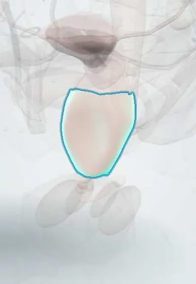
The vagina is the muscular canal from the cervix to the vulva; it is the birth canal and the passage for menstrual flow and sex.
Opens in the Durgical app, no sign-up needed.
What it is
A muscular, elastic canal about 7-10 cm long from the cervix to the vestibule, between the bladder and urethra in front and the rectum behind.
What it does
The birth canal and the passage for menstrual flow and sex; its lining is kept healthy by oestrogen and normal bacteria.
Key facts
- Where
- Fibromuscular canal 7–10 cm long from the vestibule up and back to the cervix; urethra and bladder in front, rectum and rectouterine pouch behind
- Blood supply
- Vaginal artery, vaginal branches of uterine artery, internal pudendal and middle rectal arteries
- Nerve
- Upper vagina: uterovaginal plexus (visceral); lower fifth: pudendal nerve (somatic, sensitive)
- Function
- Birth canal, menstrual outflow, sexual intercourse; lymph: upper to iliac nodes, lower to superficial inguinal nodes
Relations
- In front
- Base of the urinary bladder above and the female urethra below, embedded in its lower anterior wall, with the pubocervical fascia between
- Behind
- Upper part: rectouterine pouch; middle: rectum, across the rectovaginal septum; lower: perineal body and anal canal
- Lateral
- Levator ani (pubovaginalis), the ureters near the lateral fornices, the vaginal venous plexus and, at the opening, the vestibular bulbs and Bartholin glands
- Above
- Cervix, entering its upper anterior wall, ringed by the vaginal fornix
- Below
- Opens into the vestibule behind the urethral opening, at the hymen
How to find it
On speculum examination the blades are passed along the posterior wall, angled down and back toward the sacrum to follow the vaginal axis, then opened to show the cervix; the walls are inspected as the speculum is withdrawn. Asking the person to bear down shows any descent of the walls.
Examination
- POP-Q (Pelvic Organ Prolapse Quantification): fixed points on the anterior and posterior walls and the cervix are measured against the hymen while straining, to stage prolapse
- Single-blade (Sims) speculum: holding back one wall shows the other during straining; a front bulge means a cystocele, a back bulge a rectocele
- Vaginal pH and amine (whiff) test: a pH above 4.5 and a fishy odour when potassium hydroxide is added support bacterial vaginosis
On imaging
On sagittal T2 MRI the wall is low signal with bright mucosa and mucus inside; vaginal gel distends it to show lesions. MRI is preferred for staging vaginal and cervical cancer and for congenital anomalies such as septa or agenesis. Dynamic MRI or transperineal ultrasound on straining show prolapse of the walls. Fistulas to the bladder or rectum are shown by MRI or contrast CT.
Common problems
- Infections (vaginosis, yeast).
- Prolapse of the front or back wall (cystocele, rectocele) or of the top (vault).
- Dryness after menopause (genitourinary syndrome of menopause).
How it is diagnosed
Examination with a speculum; swabs; pelvic floor examination.
Treatment
- Treatment of infections; local oestrogen for dryness.
- Pessary or surgery for prolapse.
Clinical notes
- Vesicovaginal and rectovaginal fistulas follow prolonged obstructed labour, pelvic surgery or radiotherapy
- Vault prolapse after hysterectomy reflects loss of level I support; repairs suspend the vault from the uterosacral ligament, the sacrospinous ligament or the sacrum (sacrocolpopexy)
- Vaginal and perineal tears at birth are graded by depth, up to involvement of the anal sphincter
Anatomical variants
- A longitudinal septum dividing the vagina in two, often found with a double uterus (uterus didelphys) when the paramesonephric ducts fail to fuse
- Vaginal agenesis with an absent or rudimentary uterus (Mayer-Rokitansky-Küster-Hauser syndrome); the ovaries and external genitalia are normal
Development
The upper vagina comes from the paramesonephric ducts and the lower part from the urogenital sinus; failed canalisation gives a transverse vaginal septum, and an imperforate hymen traps menstrual blood (haematocolpos).
Related conditions
- Human papillomavirus (HPV) infection
- Bacterial vaginosis
- Vulvovaginal candidiasis
- Trichomoniasis
- Pelvic organ prolapse
- Obstetric perineal tears and anal sphincter injury
- Genital herpes (herpes simplex infection)
- Toxic shock syndrome
- Premature rupture of membranes (PROM and PPROM)
- Umbilical cord prolapse
- Sexual violence (care of the survivor)
- Uterine inversion
- Müllerian anomalies (MRKH, septate uterus, imperforate hymen)
- Genitourinary syndrome of menopause (atrophic vaginitis)
- Obstetric fistula (vesicovaginal and rectovaginal)
Test yourself
Think of the answer, then tap the question to see it.
What are the three DeLancey levels of vaginal support?
I: upper vagina hung from the cardinal and uterosacral ligaments; II: mid-vagina attached to the pelvic side wall; III: lower vagina fused to the perineal body.
Why is the posterior wall of the vagina longer than the anterior wall?
The cervix enters through the upper anterior wall.
Which findings support bacterial vaginosis on vaginal testing?
A vaginal pH above 4.5 and a fishy odour when potassium hydroxide is added (whiff test).
On a single-blade speculum examination with straining, what do anterior and posterior vaginal bulges mean?
A front bulge is a cystocele; a back bulge is a rectocele.
What does vault prolapse after hysterectomy reflect?
Loss of level I support of the vagina; repairs suspend the vault from the uterosacral, sacrospinous ligament or sacrum.
Educational information, not medical advice. For any symptom, see a health professional.
Its names
- Official Latin (TA2)
- vagina TA2 3523
- Official Spanish (DeCS)
- Vagina DeCS D014621
- English (TA2)
- Vagina
Related
Sources: Terminologia Anatomica (TA2); DeCS; Anatomy, Abdomen and Pelvis: Female Internal Genitals.
Spotted a mistake?We are always improving for the community. Tell us what is wrong and our team will review it.Report a mistake
Representative model, for educational purposes only.