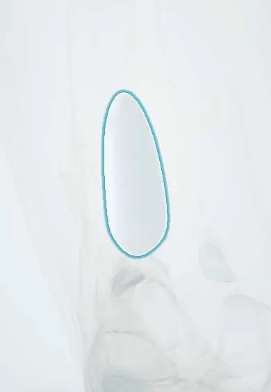
Suprapatellar bursa !
The suprapatellar bursa is a fluid sac above the kneecap that is part of the knee joint space; it fills when the knee swells.
Opens in the Durgical app, no sign-up needed.
What it is
A fluid sac above the kneecap, connected to the joint.
What it does
Lets the quadriceps tendon glide; it fills with fluid when the knee is swollen.
Key facts
- Where
- Between quadriceps tendon and femur, extending several cm above patella
- Attaches
- Held up by articularis genus muscle
- Function
- Communicates with knee joint cavity; lets quadriceps glide over femur; fills in knee effusion
Relations
- In front
- Quadriceps tendon, with the suprapatellar (quadriceps) fat pad between them at its lower end
- Behind
- Prefemoral fat pad and the front of the lower femoral shaft above the trochlea
- Above
- Articularis genus, attached to its upper end
- Below
- Opens into the knee joint above the patella and the femoral trochlea
How to find it
With the knee straight and relaxed, swelling above and on both sides of the patella that fills the normal hollows of the lower thigh marks a distended bursa. Pressing down from above the patella pushes this fluid under the kneecap, the first step of the patellar tap.
Examination
- Patellar tap: the examiner's hand first presses fluid down out of the bursa, then pushes the patella back; a tap against the femur shows a moderate effusion
On imaging
Lateral knee radiograph: an effusion makes a rounded soft-tissue density above the patella, pushing the quadriceps fat pad and the prefemoral fat pad apart. Ultrasound with the knee slightly bent and the probe along the quadriceps tendon shows small amounts of fluid, synovial thickening and loose bodies in the recess.
Clinical notes
- Effusion, haemarthrosis and septic arthritis all distend it, so swelling above the kneecap is often the first visible sign of joint fluid
- Suprapatellar plica: a remnant of the fetal partition can thicken and catch during movement, causing anterior knee pain
- A wound just above the patella can enter the joint through the bursa; the suprapatellar approach for tibial nailing also passes through it
Anatomical variants
- A suprapatellar plica (complete, partial or perforated) is found in many knees; a complete septum that cuts the bursa off from the joint is rare
Development
It starts as a sac separate from the joint; the wall between them normally breaks down before birth, and what remains forms the suprapatellar plica or, rarely, a closed bursa.
Test yourself
Think of the answer, then tap the question to see it.
Does the suprapatellar bursa communicate with the knee joint?
Yes, in most adults it opens widely into the knee and acts as part of the joint cavity, so effusion, blood or pus fill it.
Which muscle holds up the suprapatellar bursa, and why?
The articularis genus pulls it up as the knee straightens, so it is not pinched between the quadriceps tendon and the femur.
Between which structures does the suprapatellar bursa lie?
Between the quadriceps tendon in front and the femur behind, extending several centimetres above the patella.
Educational information, not medical advice. For any symptom, see a health professional.
Its names
- Official Latin (TA2)
- bursa suprapatellaris TA2 2738
- Official Spanish (DeCS)
- Bolsa suprarrotuliana DeCS D002061
- English (TA2)
- Suprapatellar bursa
Related
Sources: Terminologia Anatomica (TA2); DeCS; Anatomy, Skin Bursa; Anatomy, Bony Pelvis and Lower Limb, Knee.
Spotted a mistake?We are always improving for the community. Tell us what is wrong and our team will review it.Report a mistake
Representative model, for educational purposes only.