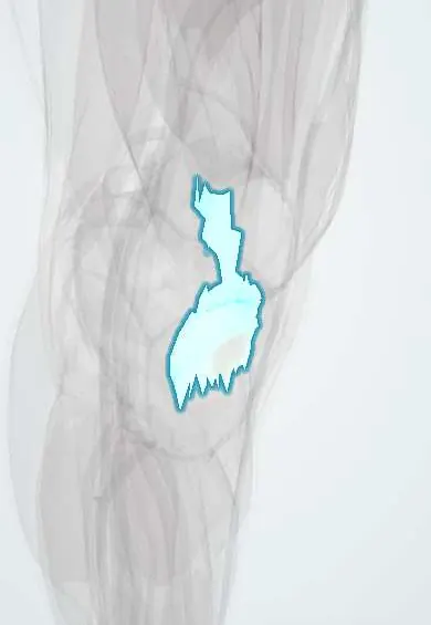
Rectus femoris tendon !
The front (superficial) layer of the quadriceps tendon above the kneecap, formed by the rectus femoris; it carries that muscle's pull to straighten the knee.
Official name: Tendon of rectus femoris
Opens in the Durgical app, no sign-up needed.
What it is
The tendon of the rectus femoris: the most superficial layer of the quadriceps tendon, running into the top of the kneecap.
What it does
Transmits the force of the quadriceps to straighten the knee.
Relations
- Above
- The rectus femoris belly, whose lower aponeurosis narrows into this tendon in the lower thigh
- Superficial
- Fascia lata and skin; over the patella, the prepatellar bursa lies in front of its fibres
- Deep
- The merged tendons of the vastus medialis and vastus lateralis, then the vastus intermedius tendon and the suprapatellar bursa
- Medial
- Vastus medialis and its expansion to the medial retinaculum
- Lateral
- Vastus lateralis and its expansion to the lateral retinaculum
- Below
- Base and front of the patella and, through its superficial fibres, the patellar tendon
How to find it
With the knee straight, the person tightens the thigh: a firm, flat cord is felt in the midline just above the base of the patella, continuous above with the rectus femoris belly.
Examination
- Ely test: prone, the knee is bent passively; if the hip flexes and the buttock rises, the rectus femoris unit is tight
On imaging
Ultrasound and MRI show the quadriceps tendon as three to four stacked layers; in a partial tear the superficial (rectus femoris) layer is often the one disrupted.
Common problems
- Tendinitis from repeated jumping or kneeling.
- Rupture, usually in older adults; the leg cannot be straightened.
How it is diagnosed
Physical exam; ultrasound or MRI.
Treatment
- Tendinitis: rest, physical therapy.
- Complete tear: surgical repair, usually soon after the injury.
- Part of this tendon can also be used as an ACL graft.
Clinical notes
- In a partial quadriceps tendon rupture the rectus femoris layer may tear while the deeper vasti layers still allow weak extension, so the injury can be missed
- Quadriceps tendon grafts for ACL reconstruction are taken from the central strip, which includes this superficial layer, with or without a block of patellar bone
Test yourself
Think of the answer, then tap the question to see it.
Which quadriceps muscle forms the most superficial layer of the quadriceps tendon?
The rectus femoris: its tendon lies in front of the merged vasti and the deep vastus intermedius tendon.
Where do the superficial fibres of the rectus femoris tendon end?
They pass over the front of the patella into the patellar tendon and so reach the tibial tuberosity.
Why is the rectus femoris unit stretched most with the hip extended and the knee bent?
It is the only quadriceps head that crosses the hip, so it lengthens across both joints in that position.
Why can a partial quadriceps tendon tear limited to the rectus femoris layer be missed?
The deeper vasti layers stay intact and still allow weak active knee extension.
Educational information, not medical advice. For any symptom, see a health professional.
Its names
- Official Latin (TA2)
- tendo musculi quadricipitis femoris TA2 2621
- Official Spanish (DeCS)
- Tendón del músculo recto femoral DeCS D013710
- English (TA2)
- Tendon of rectus femoris
Related
Sources: Terminologia Anatomica (TA2); DeCS; Anatomía humana (4.ª ed.), tomo 1, cap. 65: Rodilla. Músculos del muslo; Anatomy, Bony Pelvis and Lower Limb: Thigh Quadriceps Muscle.
Spotted a mistake?We are always improving for the community. Tell us what is wrong and our team will review it.Report a mistake
Representative model, for educational purposes only.