Joints and ligaments · Lower limb
Joint capsule !
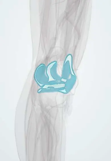
The knee joint capsule is a sleeve around the knee, lined inside with a membrane that makes the joint fluid; swelling fills it.
Official name: Articular capsule of knee joint
Opens in the Durgical app, no sign-up needed.
What it is
A sleeve of tissue around the whole joint, lined inside by synovium.
What it does
Encloses the joint and holds the joint fluid that lubricates it.
Key facts
- Where
- Encloses knee joint; replaced anteriorly by patella, quadriceps tendon and patellar ligament
- Attaches
- Articular margins of femur and tibia; posteriorly above condyles and intercondylar fossa
- Function
- Holds synovial fluid; largest synovial cavity, communicates with suprapatellar bursa; reinforced by retinacula and popliteal ligaments; genicular nerves and arterial anastomosis; flexion 140–160°; rotation only when bent (lateral ~35°, medial ~10°)
Relations
- In front
- Quadriceps tendon, patella and patellar ligament take its place in front, with the medial and lateral patellar retinacula on either side; the infrapatellar fat pad lies inside it, behind the patellar ligament
- Behind
- Oblique and arcuate popliteal ligaments, the two heads of gastrocnemius and the popliteus; the popliteal artery lies directly on the capsule, the deepest structure of the popliteal fossa
- Medial
- The deep part of the medial collateral ligament is a thickening of the capsule and is bound to the medial meniscus; the superficial MCL and the pes anserinus tendons lie further out
- Lateral
- The lateral collateral ligament stays separate from the capsule; the popliteus tendon runs between the two, inside the capsule, separating the lateral meniscus from the LCL
- Above
- Continues as the suprapatellar bursa beneath the quadriceps tendon, its apex held up by articularis genus
- Contents
- Synovial cavity with the articular cartilage and the menisci; the cruciate ligaments and the infrapatellar fat pad sit within the fibrous layer but outside the synovium
How to find it
With the knee bent to about 90°, the soft hollows beside the patellar ligament lead to the edges of the tibial plateau (joint line). With the knee straight, the patellar apex lies about 1 cm above that line (some texts: at its level); the fibular head is felt posterolaterally about two finger-breadths below it. An effusion, not the capsule, is felt; it is aspirated via the suprapatellar recess.
Examination
- Bulge (sweep) test: fluid stroked up out of the medial gutter returns as a bulge on the medial side when the lateral side is stroked; it detects small effusions
- Patellar tap (ballottement): with the suprapatellar pouch pressed empty, pushing the patella down makes it tap on the femur; positive with moderate or large effusions
On imaging
Lateral radiograph: an effusion fills the suprapatellar recess as a soft-tissue density between the quadriceps and prefemoral fat pads; a fat–fluid level on a horizontal-beam view (lipohaemarthrosis) points to an intra-articular fracture. Ultrasound shows fluid and synovial thickening and guides needles; MRI shows synovitis, capsular tears and loose bodies.
Clinical notes
- Septic arthritis: pus fills the whole synovial cavity including the suprapatellar recess; the posterior communication with the semimembranosus–gastrocnemius bursa can carry fluid into a popliteal cyst
- A slit-like, often one-way opening in the posteromedial capsule lets joint fluid collect behind the knee as a Baker (popliteal) cyst; joint disease such as osteoarthritis or a meniscal tear usually drives it
- Crystal arthritis is confirmed in fluid drawn from the capsule: needle-shaped, negatively birefringent urate crystals in gout; rhomboid, weakly positively birefringent calcium pyrophosphate crystals in pseudogout
- An effusion reflexly inhibits the quadriceps, especially vastus medialis, which is why a swollen knee quickly loses extension strength
Anatomical variants
- Synovial plicae, remnants of embryonic partitions: suprapatellar, medial patellar and infrapatellar (ligamentum mucosum); a thick medial plica can catch on the medial femoral condyle and cause anterior knee pain
Development
From week 6 the knee forms between the cartilage models of the femur, tibia and patella: the middle of the interzone breaks down into the cavity, the mesenchyme around it becomes the capsule and ligaments, and part of the interzone stays as the menisci. Partitions of the early cavity that fail to regress remain as plicae.
Related conditions
- Baker's (Popliteal) Cyst
- Septic Arthritis of the Knee
- Gout of the Knee
- Pseudogout (CPPD) of the Knee
- Chikungunya
- Gout
- Haemophilia A and B
- Reactive arthritis
- IgA vasculitis (Henoch–Schönlein purpura)
- Juvenile idiopathic arthritis
- Adult-onset Still disease
Test yourself
Think of the answer, then tap the question to see it.
Are the cruciate ligaments inside the knee capsule?
Inside the fibrous capsule but outside the synovial cavity: intracapsular and extrasynovial.
Why does a knee effusion first show as fullness above and beside the patella?
The joint cavity runs up above the patella as the suprapatellar bursa, which fills first.
Which bedside tests detect a knee effusion?
The bulge (sweep) test for small effusions, and the patellar tap (ballottement) for moderate or large ones.
What does a fat–fluid level on a horizontal-beam lateral knee radiograph indicate?
Lipohaemarthrosis, pointing to an intra-articular fracture.
Which crystals in knee fluid confirm gout and which pseudogout?
Gout: needle-shaped, negatively birefringent urate crystals. Pseudogout: rhomboid, weakly positively birefringent calcium pyrophosphate crystals.
Educational information, not medical advice. For any symptom, see a health professional.
Its names
- Official Latin (TA2)
- articulatio genus; capsula articularis TA2 1883
- Official Spanish (DeCS)
- Cápsula articular de la rodilla DeCS D017746
- English (TA2)
- Articular capsule of knee joint
- Also called
- articulus genus
Related
Sources: Terminologia Anatomica (TA2); DeCS; Anatomy, Bony Pelvis and Lower Limb, Knee; 9.7 Development of Joints (OpenStax (Rice University), 2022) · adapted, CC BY 4.0; Anatomy of the Human Body (20th ed.): Development of the Joints (Lea & Febiger (Gray's Anatomy, 1918), 1918) · adapted, Public domain; Anatomía humana (4.ª ed.), tomo 1, cap. 65: Rodilla. Músculos del muslo.
Spotted a mistake?We are always improving for the community. Tell us what is wrong and our team will review it.Report a mistake
Representative model, for educational purposes only.