Digestive system · Head and neck
Upper right lateral incisor !
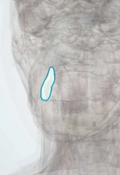
The upper right lateral incisor (tooth 12) is the flat front tooth next to the central incisor; it helps cut food.
Opens in the Durgical app, no sign-up needed.
What it is
A front tooth beside the central incisor, a little smaller, with a cutting edge and one root.
What it does
Bites into and cuts food.
Key facts
- Joints with
- Alveolar socket of the maxilla (gomphosis held by the periodontal ligament); bites against the opposing teeth
- Landmarks
- FDI 12, upper right lateral incisor: 1 root; smaller than the central incisor; often small, peg-shaped or missing; erupts at about 8–9 years; nerve: anterior superior alveolar nerve (V2)
Relations
- Medial
- The upper right central incisor
- Lateral
- The upper right canine; in children the crown of the developing canine lies close to this tooth's root
- In front
- Upper lip, the orbicularis oris and the labial gum
- Below
- Opposes the lower right lateral incisor and the lower right canine
How to find it
Second tooth from the midline on the upper right; narrower and shorter than the central incisor beside it, with a more rounded edge.
On imaging
On a periapical, dens invaginatus shows as a loop of enamel folding into the crown toward the pulp. A panoramic film confirms a missing lateral and shows where the canine above it is heading.
Common problems
- Sometimes small or missing from birth (more often the upper one).
Clinical notes
- Anterior superior alveolar nerve (V2); infiltration in the vestibule above the apex numbs it, and the nasopalatine nerve also reaches the palatal gum here
- Its root tip often curves backward and leans toward the palate, so an abscess from this tooth may point on the palate rather than in the vestibule
Anatomical variants
- Among the teeth most often missing from birth; a small cone-shaped (peg) crown is a milder form of the same tendency
Development
Replaces the primary upper right lateral incisor (FDI 52).
Related conditions
Test yourself
Think of the answer, then tap the question to see it.
Which developmental anomaly most often affects the upper right lateral incisor?
Dens invaginatus: an enamel fold that dips toward the pulp, linked to the pit on its palatal surface.
What happens to the upper right canine when the upper right lateral incisor is peg-shaped or missing?
The canine loses its eruption guide (the lateral incisor's root) and tends to erupt toward the palate.
Why may an abscess from the upper right lateral incisor point on the palate?
Its root tip often curves backward and leans toward the palate.
How does dens invaginatus of the upper right lateral incisor look on a periapical radiograph?
As a loop of enamel folding from the palatal pit into the crown toward the pulp.
Educational information, not medical advice. For any symptom, see a health professional.
Related
Sources: Anatomy, Head and Neck, Teeth; Anatomy, Permanent Dentition; Anatomy, Head and Neck, Tooth Eruption; Anatomy, Head and Neck: Alveolar Nerve; 23.3 The Mouth, Pharynx, and Esophagus (OpenStax (Rice University), 2022) · adapted, CC BY 4.0.
Spotted a mistake?We are always improving for the community. Tell us what is wrong and our team will review it.Report a mistake
Representative model, for educational purposes only.