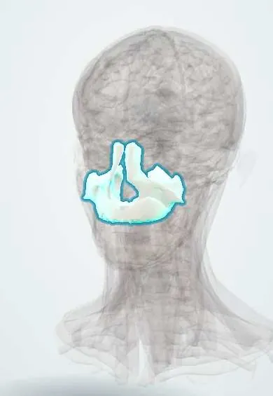
Skeletal system · Head and neck
Maxillae !
The maxillae are the two bones of the upper jaw; they hold the upper teeth and the maxillary sinuses and form the middle of the face and most of the palate.
Official name: Maxilla
Opens in the Durgical app, no sign-up needed.
What it is
The two bones of the upper jaw; they hold the upper teeth and the maxillary sinuses.
What it does
Form the middle of the face, the floor of the eye sockets and most of the hard palate.
Key facts
- Joints with
- Opposite maxilla, frontal, nasal, lacrimal, ethmoid, zygomatic, palatine, vomer, inferior concha; upper teeth
- Landmarks
- Maxillary sinus, infraorbital foramen, alveolar process, palatine process, frontal process; infraorbital groove and canal; incisive canal (nasopalatine nerve); maxillary tuberosity (posterior superior alveolar nerves); canine fossa; anterior nasal spine
Relations
- Above
- Orbit: the orbital floor with the inferior rectus and inferior oblique just above it
- Medial
- Nasal cavity: the inferior nasal concha and the inferior meatus with the nasolacrimal duct opening; the opposite maxilla at the intermaxillary suture
- Lateral
- Zygomatic bone at the zygomatic process
- Behind
- Infratemporal and pterygopalatine fossae, with the maxillary artery and the posterior superior alveolar nerves on the maxillary tuberosity
- Below
- Upper teeth in the alveolar process; the mouth below the palatine process
- Superficial
- Levator labii superioris and levator anguli oris; the infraorbital nerve emerging from the infraorbital foramen
How to find it
The infraorbital rim and the front of the maxilla are felt below the eye; the infraorbital foramen lies about 1 cm below the rim, roughly in line with the pupil.
Examination
- Midface mobility: with the forehead steadied, the upper teeth and palate are rocked; the palate moving alone suggests Le Fort I, with the nasal bridge Le Fort II, the whole face Le Fort III
- Forced duction test: restricted passive movement of the globe suggests mechanical entrapment in an orbital floor fracture rather than a nerve palsy
On imaging
Facial CT with coronal reconstructions is the standard for midface and orbital floor fractures (look for orbital tissue hanging into the sinus, the «teardrop», and blood in the maxillary sinus); the occipitomental (Waters) radiograph shows the orbital rims and sinuses.
Clinical notes
- In children an orbital floor «trapdoor» fracture can trap the inferior rectus with little bruising (white-eyed blowout), with nausea and a slow pulse from the oculocardiac reflex; it is treated as urgent
- Infraorbital nerve block at the foramen numbs the lower eyelid, side of the nose, cheek and upper lip
- Le Fort II and III fractures can tear the dura at the skull base, so CSF rhinorrhoea is looked for
Anatomical variants
- Torus palatinus: a smooth, hard bony bump in the midline of the hard palate; common and harmless, though it can interfere with an upper denture
- Accessory infraorbital foramina in a minority of people; they can carry nerve branches that a block at the main foramen does not reach
Development
Forms in membrane from the maxillary prominence of the first pharyngeal arch; failure to fuse with the medial nasal prominence gives cleft lip, and failure of the palatal shelves to fuse gives cleft palate.
Related conditions
- Periodontitis
- Cleft lip and palate
- Melasma
- Fibrous dysplasia of bone and McCune–Albright syndrome
- Midface fractures (Le Fort, zygomatic, nasal)
Test yourself
Think of the answer, then tap the question to see it.
How do Le Fort I, II and III fractures of the maxilla differ?
I: horizontal above the teeth (mobile palate). II: pyramidal through the nasal bridge and orbital floors. III: separates the face from the skull.
What are the signs of a blowout fracture of the orbital floor of the maxilla?
Double vision on upward gaze from inferior rectus entrapment, a sunken eye (enophthalmos) and a numb cheek from infraorbital nerve injury.
Why is the paediatric «white-eyed» orbital floor fracture of the maxilla treated as urgent?
A trapdoor fracture traps the inferior rectus with little bruising, causing nausea and a slow pulse from the oculocardiac reflex.
Which test distinguishes mechanical entrapment in an orbital floor fracture from a nerve palsy?
The forced duction test: restricted passive movement of the globe suggests mechanical entrapment.
What complication can follow extraction of upper molars whose roots project into the maxillary sinus?
An oroantral communication between the mouth and the maxillary sinus.
Educational information, not medical advice. For any symptom, see a health professional.
Its names
- Official Latin (TA2)
- maxilla TA2 756
- Official Spanish (DeCS)
- Maxilar DeCS D008437
- English (TA2)
- Maxilla
- Also called
- os maxillare · maxillary bone
Related
- Maxillary sinus
- Upper right central incisor
- Upper right lateral incisor
- Upper right canine
- Upper right first premolar
- Upper right second premolar
- Upper right first molar
- Upper right second molar
Sources: Terminologia Anatomica (TA2); DeCS; Anatomy, Head and Neck, Maxilla.
Spotted a mistake?We are always improving for the community. Tell us what is wrong and our team will review it.Report a mistake
Representative model, for educational purposes only.