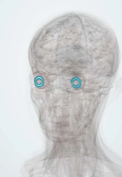
Zonular fibres !
The zonular fibres are hundreds of fine threads from the ciliary body to the edge of the lens; they hold the lens in place and pass on the pull that focuses it.
Official name: Ciliary zonule
Opens in the Durgical app, no sign-up needed.
What it is
Hundreds of fine fibres from the ciliary body to the edge of the lens.
What it does
Hold the lens in place and pass the ciliary muscle's pull to it.
Key facts
- Attaches
- Ciliary processes to the lens capsule equator
- Function
- Suspend the lens; transmit ciliary muscle relaxation
- Tight in
- Distance vision (ciliary muscle relaxed): lens flattened
Relations
- Course
- From the pars plana and the valleys between the ciliary processes, forward and inward to the lens capsule, attaching in front of, at and behind its equator
- In front
- Posterior chamber and, beyond it, the back of the iris; aqueous flows between the fibres
- Behind
- Front face of the vitreous body
How to find it
Not seen in a normal eye, as they lie behind the iris. They show at the slit lamp through a dilated pupil when the lens is displaced, or on ultrasound biomicroscopy.
Examination
- Slit lamp: a lens tremor (phacodonesis) or iris tremor (iridodonesis) on eye movement points to zonular weakness
- Dilated exam: the lens edge seen within the pupil means zonular breaks (subluxation)
On imaging
Ultrasound biomicroscopy shows the zonules and any gaps; B-scan ultrasound finds a lens fully dislocated into the vitreous.
Clinical notes
- Pseudoexfoliation syndrome deposits flaky material on the lens and zonules: weak zonules plus a higher risk of glaucoma
- In cataract surgery a capsular tension ring can support a lens bag with partial zonular loss
- Blunt trauma can tear zonules (zonular dialysis), tilting or dislocating the lens
Development
Laid down as the tertiary vitreous, secreted by the ciliary epithelium.
Memory aid
Close-up: Contract, slacken, round (the ciliary muscle contracts, the zonules go slack, the lens rounds).
Related conditions
Test yourself
Think of the answer, then tap the question to see it.
Which protein makes up the zonular fibres, and why does the lens dislocate in Marfan syndrome?
Fibrillin-1, the protein affected in Marfan syndrome, so the zonules are weak.
Are the zonular fibres taut or slack for distance vision, and what happens to the lens?
Taut: the ciliary muscle relaxes, the zonules tighten and flatten the lens (Helmholtz theory).
Which slit-lamp signs point to zonular weakness?
Phacodonesis (lens tremor) or iridodonesis (iris tremor) on eye movement.
Educational information, not medical advice. For any symptom, see a health professional.
Its names
- Official Latin (TA2)
- zonula ciliaris TA2 6795
- Official Spanish (DeCS)
- Fibras zonulares
- English (TA2)
- Ciliary zonule
- Also called
- apparatus suspensorius lentis · zonula Zinni · zonule of Zinn
Related
Sources: Terminologia Anatomica (TA2); Anatomy, Head and Neck, Eye Ciliary Muscles; Anatomy, Head and Neck, Eye.
Spotted a mistake?We are always improving for the community. Tell us what is wrong and our team will review it.Report a mistake
Representative model, for educational purposes only.