Vitreous !
The vitreous is the clear gel filling the back of the eye; it keeps the eye's shape, and clumps in it cast the shadows called floaters.
Official name: Vitreous body
Opens in the Durgical app, no sign-up needed.
What it is
The clear gel filling the back of the eye.
What it does
Keeps the eye's shape; clumps in it cast the shadows called floaters.
Key facts
- Where
- Fills the space between the lens and the retina (~80% of the eye)
- Blood supply
- Avascular
- Function
- Keeps the eye's shape; transparent path for light
Relations
- In front
- Back of the lens, which sits in its hyaloid fossa, and the zonular fibres
- Behind
- Optic disc and macula, where the posterior hyaloid face adheres
- Superficial
- Retina, against the internal limiting membrane, and the pars plana of the ciliary body at its base
- Contents
- Cloquet canal, the path of the former hyaloid artery from disc to lens, and scattered cells (hyalocytes)
How to find it
Seen at the slit lamp just behind the lens (anterior vitreous) and with the ophthalmoscope as a clear space; floaters and pigment drift in it as the eye moves.
Examination
- Slit lamp of the anterior vitreous: pigment cells (tobacco dust, Shafer sign) after a posterior vitreous detachment strongly suggest a retinal tear
- Fundoscopy: a floating ring in front of the disc (Weiss ring) marks a posterior vitreous detachment
On imaging
B-scan ultrasound shows the detached posterior hyaloid, and any retinal detachment, when blood blocks the view; OCT shows vitreomacular traction.
Common problems
- Floaters: specks and cobwebs from clumping gel, common with age.
- Posterior vitreous detachment: the gel peels off the retina; a sudden burst of floaters needs a retina check.
- Vitreous haemorrhage, often from diabetic vessels.
How it is diagnosed
Dilated eye exam; ultrasound when blood blocks the view.
Treatment
- Usually no treatment for floaters.
- Vitrectomy (removing the gel) for persistent bleeding or retinal problems.
Clinical notes
- Injections into the vitreous (anti-VEGF drugs, steroids, antibiotics) and vitrectomy instruments enter through the pars plana
- Endophthalmitis: infection of the vitreous after surgery or injury; a sample is taken from it for culture
- Asteroid hyalosis: many small white calcium-lipid particles, usually harmless
- Persistent fetal vasculature (persistent hyperplastic primary vitreous) causes leukocoria and a small eye in infants
Anatomical variants
- Remnants of the fetal hyaloid artery are common and harmless: a small dot on the back of the lens capsule (Mittendorf dot) or a tuft of glial tissue on the disc (Bergmeister papilla)
Development
The primary vitreous, which holds the hyaloid vessels, is replaced by the avascular secondary vitreous; the tertiary vitreous forms the zonular fibres.
Related conditions
Test yourself
Think of the answer, then tap the question to see it.
Why are new flashes and floaters checked with a dilated retinal exam?
They may mean a posterior vitreous detachment that has torn the retina, which can lead to retinal detachment.
Where is the vitreous body attached most firmly?
At the vitreous base over the ora serrata; less so at the optic disc, the macula and along vessels.
What does «tobacco dust» (Shafer sign) in the anterior vitreous suggest?
A retinal tear, after a posterior vitreous detachment.
What is Terson syndrome?
Bleeding into the eye (vitreous haemorrhage) with a subarachnoid haemorrhage.
Educational information, not medical advice. For any symptom, see a health professional.
Its names
- Official Latin (TA2)
- corpus vitreum TA2 6809
- Official Spanish (DeCS)
- Cuerpo Vítreo DeCS D014822
- English (TA2)
- Vitreous body
- Also called
- vitreous humour
Related
Sources: Terminologia Anatomica (TA2); DeCS; Posterior Vitreous Detachment; Anatomy, Head and Neck, Eye.
Spotted a mistake?We are always improving for the community. Tell us what is wrong and our team will review it.Report a mistake
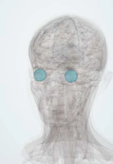
Representative model, for educational purposes only.