Lens !
The lens is a clear, flexible disc behind the iris; it fine-tunes focus for near and far, and it turns cloudy in a cataract.
Opens in the Durgical app, no sign-up needed.
What it is
A clear, flexible disc behind the iris, hung by the zonular fibres.
What it does
Fine-tunes focus for near and far; it clouds in cataract.
Key facts
- Where
- Behind the iris, hung by the zonules
- Blood supply
- Avascular (aqueous humour)
- Function
- Fine focus (accommodation, up to ~10–15 D when young); clouds in cataract
Relations
- In front
- Iris, which rests on it at the pupil margin, and the posterior chamber
- Behind
- Vitreous body, in a shallow hollow on the vitreous face (hyaloid fossa)
- Superficial
- At its equator, the zonular fibres and, beyond them, the ciliary processes of the ciliary body
How to find it
Behind the pupil and invisible in a healthy eye; opacities show as dark shadows against the red reflex. It is examined at the slit lamp after dilating the pupil.
Examination
- Red reflex with an ophthalmoscope at arm's length: dark spots suggest cataract; a white reflex (leukocoria) in a child points to congenital cataract or retinoblastoma and is an urgent finding
- Near point of accommodation: it recedes with age, which is presbyopia
- Slit lamp with retroillumination: grades nuclear, cortical (spoke-like) and posterior subcapsular cataract
On imaging
Optical or ultrasound biometry measures eye length and corneal power to choose the artificial lens; B-scan ultrasound finds a lens dislocated into the vitreous and shows the retina behind a dense cataract.
Common problems
- Cataract: the lens clouds slowly with age (also diabetes, steroids, injury), making vision blurry and glary.
- Presbyopia: after about 45 the lens stiffens, so reading up close gets hard.
How it is diagnosed
Vision test and slit-lamp exam with the pupil dilated.
Treatment
- Reading glasses for presbyopia; brighter light and updated glasses for early cataract.
- Cataract surgery: the cloudy lens is broken up and replaced by a clear artificial lens.
Clinical notes
- Cataract types: nuclear (often with a myopic shift), cortical, and posterior subcapsular (steroids, diabetes; marked glare); oil-droplet cataract in galactosaemia; congenital cataract after maternal rubella
- Cataract surgery breaks up and removes the lens contents and places an artificial lens in the capsular bag; later clouding of the back of the capsule is opened with a laser
- A swollen (intumescent) lens can close the angle, and leaking lens proteins can clog the meshwork (lens-induced glaucoma)
- A small, round lens (microspherophakia) in Weill–Marchesani syndrome can block the pupil and cause angle closure
Development
The optic vesicle induces the surface ectoderm to thicken into the lens placode, which folds into the lens vesicle; its oldest fibres form the embryonic nucleus at the centre.
Memory aid
Marfan looks up (lens up and out); homocystinuria looks down (down and in).
Related conditions
- Homocystinuria
- Classic galactosaemia
- Cataract
- Acute angle-closure glaucoma
- Refractive errors and presbyopia
- Rubella and congenital rubella syndrome
- Marfan syndrome
Test yourself
Think of the answer, then tap the question to see it.
What happens to the ciliary muscle, the zonular fibres and the lens during accommodation for near?
The ciliary muscle contracts, the zonular fibres slacken and the elastic capsule rounds the lens; part of the near triad with convergence and miosis.
In which direction does the lens usually dislocate in Marfan syndrome and in homocystinuria?
Marfan syndrome: up and out; homocystinuria: down and in.
What does a white pupillary reflex (leukocoria) in a child suggest?
Congenital cataract or retinoblastoma: an urgent finding.
What does cataract surgery do, and how is later clouding of the capsule treated?
It breaks up and removes the lens contents and places an artificial lens in the capsular bag; later posterior capsule clouding is opened with a laser.
Educational information, not medical advice. For any symptom, see a health professional.
Its names
- Official Latin (TA2)
- lens TA2 6798
- Official Spanish (DeCS)
- Cristalino
- English (TA2)
- Lens
- Also called
- crystalline lens · lens crystallina
Related
Sources: Terminologia Anatomica (TA2); Ectopia Lentis; Anatomy, Head and Neck, Eye.
Spotted a mistake?We are always improving for the community. Tell us what is wrong and our team will review it.Report a mistake
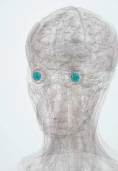
Representative model, for educational purposes only.