Ciliary body !
The ciliary body is a ring of muscle and folds behind the iris; it changes the shape of the lens to focus and makes the fluid inside the eye.
Opens in the Durgical app, no sign-up needed.
What it is
A ring of muscle and folds behind the iris.
What it does
Changes the lens shape to focus and makes the aqueous humour.
Key facts
- Where
- Ring behind the iris, from the iris root to the ora serrata
- Blood supply
- Long posterior and anterior ciliary arteries
- Nerve
- Ciliary muscle: parasympathetic (III via short ciliary nerves)
- Function
- Accommodation; makes aqueous humour (ciliary processes)
Relations
- In front
- Iris root and the drainage angle; on gonioscopy its front face is the ciliary body band, between the iris root and the scleral spur
- Behind
- Choroid and retina, which it meets at the ora serrata
- Superficial
- Sclera, with the longitudinal fibres of the ciliary muscle anchored to the scleral spur
- Deep
- Posterior chamber, the zonular fibres running to the lens equator, and the vitreous base over the pars plana
How to find it
Not visible from outside: it lies under the sclera as a ring a few millimetres behind the limbus. It is seen on gonioscopy as the ciliary body band and in full on ultrasound biomicroscopy.
Examination
- Accommodation (near point) testing: poor near focus in a young person suggests a paralysed ciliary muscle, as after an oculomotor nerve palsy or cycloplegic drops
- Slit lamp: a ring of redness around the limbus (ciliary flush) points to inflammation inside the eye rather than conjunctivitis
On imaging
Ultrasound biomicroscopy shows the ciliary processes, the pars plana and cysts or tumours behind the iris; anterior segment OCT shows only its front part.
Clinical notes
- Cycloplegic drops paralyse the ciliary muscle: used for refraction in children and to ease the painful spasm of anterior uveitis
- Intermediate uveitis (pars planitis) leaves white exudates over the pars plana and the lower vitreous (snowbanks and snowballs)
- In refractory glaucoma, laser or freezing treatment of the ciliary processes (cyclodestruction) reduces aqueous production
- A ciliary body melanoma can hide behind the iris; dilated episcleral (sentinel) vessels over it are a clue
Development
Its two-layered epithelium comes from the rim of the optic cup (neuroectoderm); the ciliary muscle and stroma come from neural crest mesenchyme.
Related conditions
Test yourself
Think of the answer, then tap the question to see it.
Which part of the ciliary body secretes aqueous humour, and which enzyme do some glaucoma drugs target there?
The non-pigmented epithelium of the ciliary processes; carbonic anhydrase, whose inhibition cuts aqueous production.
What are the two parts of the ciliary body, and which one is the safe route for intravitreal injections?
The folded pars plicata in front (ciliary processes) and the flat pars plana behind, the safe entry route into the vitreous.
What is the nerve supply of the ciliary muscle?
Parasympathetic fibres from the oculomotor nerve (III) via the short ciliary nerves.
Why are cycloplegic drops used in children and in anterior uveitis?
They paralyse the ciliary muscle: for accurate refraction in children and to ease the painful ciliary spasm of anterior uveitis.
Educational information, not medical advice. For any symptom, see a health professional.
Its names
- Official Latin (TA2)
- corpus ciliare TA2 6765
- Official Spanish (DeCS)
- Cuerpo Ciliar DeCS D002924
- English (TA2)
- Ciliary body
Related
Sources: Terminologia Anatomica (TA2); DeCS; Anatomy, Head and Neck, Eye Ciliary Muscles; Uveitis.
Spotted a mistake?We are always improving for the community. Tell us what is wrong and our team will review it.Report a mistake
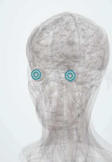
Representative model, for educational purposes only.