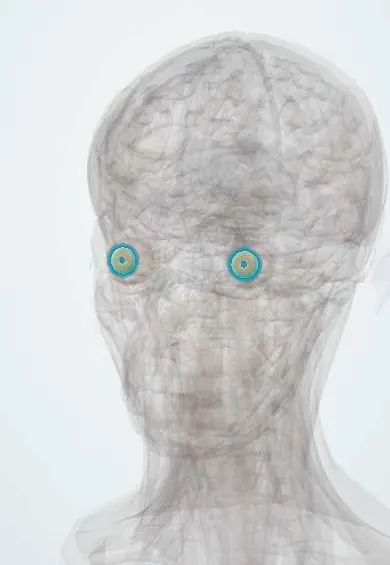
Iris !
The iris is the coloured ring of the eye; its two tiny muscles change the size of the pupil to control how much light gets in.
Opens in the Durgical app, no sign-up needed.
What it is
The coloured ring of the eye, with two tiny muscles (sphincter and dilator).
What it does
Changes the size of the pupil to control how much light gets in.
Key facts
- Where
- Between the cornea and the lens, part of the uvea
- Blood supply
- Long posterior and anterior ciliary arteries (major arterial circle)
- Nerve
- Sphincter: parasympathetic (Edinger-Westphal nucleus, III, ciliary ganglion, short ciliary nerves); dilator: sympathetic (ciliospinal centre C8–T2, superior cervical ganglion, internal carotid plexus, long ciliary nerves)
- Function
- Controls pupil size
Relations
- In front
- Anterior chamber and, beyond it, the cornea
- Behind
- Posterior chamber; the front of the lens supports its pupillary margin, and the ciliary processes lie behind its root
- Contents
- Sphincter and dilator muscles, a stroma rich in melanocytes, and the major arterial circle at its root, from which radial vessels run towards the pupil
How to find it
The coloured ring seen through the cornea, with the pupil at its centre; its root joins the ciliary body at the drainage angle. At the slit lamp its front surface shows crypts and the collarette, a ridge a short way from the pupil margin. The pupil is roughly 2–4 mm in bright light and up to about 8 mm in the dark in young adults, and it narrows with age.
Examination
- Pupil light reflex, direct and consensual: an efferent defect (oculomotor nerve or iris) leaves that pupil sluggish to both; an afferent defect (optic nerve, retina) shows as a relative afferent pupillary defect on the swinging flashlight test
- Near reflex: better constriction to near than to light (light-near dissociation) suggests Argyll Robertson pupils of neurosyphilis or an Adie tonic pupil; dilute pilocarpine constricts an Adie pupil, while a pupil blocked by atropine stays wide even with the standard strength
- Anisocoria in dim and bright light: larger difference in the dark means the small pupil is abnormal (Horner); larger in bright light means the large pupil is abnormal
- Apraclonidine or cocaine eye drops confirm Horner syndrome: the small pupil dilates with apraclonidine and fails to dilate with cocaine
On imaging
Seen directly at the slit lamp, with gonioscopy for the angle at its root. Anterior segment OCT and ultrasound biomicroscopy show its profile in cross section: a forward-bowed iris (iris bombé) or a narrow angle in angle-closure glaucoma, and the size and depth of an iris cyst or tumour. On CT or MRI of the orbit it is only a thin line in front of the lens.
Common problems
- Iritis (anterior uveitis): a painful red eye with light sensitivity.
- Unequal pupils (anisocoria): often harmless, but new unequal pupils with a droopy lid or headache need urgent care.
How it is diagnosed
Slit-lamp exam and checking how each pupil reacts to light and to near focus.
Treatment
- Steroid and pupil-dilating drops for iritis.
- Urgent scans when unequal pupils come with other nerve signs.
Clinical notes
- Anterior uveitis can stick the iris to the lens (posterior synechiae) and make the pupil irregular; if the whole margin sticks, the iris bows forward (iris bombé) and closes the angle
- Heterochromia: a lighter iris on the side of a congenital Horner syndrome; also Waardenburg syndrome, Fuchs heterochromic iridocyclitis, and darkening from prostaglandin eye drops
- Lisch nodules (pigmented iris hamartomas) in neurofibromatosis type 1; Brushfield spots in Down syndrome
- New vessels on the iris (rubeosis) in proliferative diabetic retinopathy or retinal vein occlusion herald neovascular glaucoma
- Acute angle closure leaves a mid-dilated, sluggish, often oval pupil in a red, painful eye; dilating drops for a fundus exam can rarely set it off when the angle is narrow
Anatomical variants
- Persistent pupillary membrane: fine strands across the pupil left from that fetal membrane, common and usually harmless
Development
The iris muscles come from the neuroectoderm of the optic cup, an exception among muscles; the stroma comes from neural crest. Failed closure of the optic fissure leaves an inferonasal (keyhole) coloboma. A thin vascular membrane closes the fetal pupil until shortly before birth. Aniridia (PAX6) can be part of WAGR syndrome with Wilms tumour.
Memory aid
Parasympathetic makes the pupil Pinpoint; sympathetic opens it wide for fight or flight.
Related conditions
- Oculocutaneous albinism
- Acute angle-closure glaucoma
- Uveitis
- Ocular trauma: hyphema and open globe injury
- Chédiak–Higashi syndrome
- Cataract
Test yourself
Think of the answer, then tap the question to see it.
What is the nerve supply of the sphincter pupillae and the dilator pupillae?
Sphincter: parasympathetic (Edinger–Westphal, oculomotor nerve, ciliary ganglion, short ciliary nerves); dilator: sympathetic, via the long ciliary nerves.
Why does a compressive oculomotor nerve palsy give a large unreactive pupil?
The pupil fibres run on the outside of the oculomotor nerve, so compression (posterior communicating artery aneurysm, uncal herniation) hits them first.
What are the features of Horner syndrome?
A small pupil that dilates slowly in the dark, mild ptosis and anhidrosis, from a lesion anywhere on the sympathetic path.
If anisocoria is greater in dim light, which pupil is abnormal?
The small pupil, which fails to dilate, as in Horner syndrome; a larger difference in bright light means the large pupil is abnormal.
What do new vessels on the iris (rubeosis) herald?
Neovascular glaucoma, typically from proliferative diabetic retinopathy or retinal vein occlusion.
Educational information, not medical advice. For any symptom, see a health professional.
Its names
- Official Latin (TA2)
- iris TA2 6753
- Official Spanish (DeCS)
- Iris DeCS D007498
- English (TA2)
- Iris
Related
Sources: Terminologia Anatomica (TA2); DeCS; Anatomy, Head and Neck: Eye Iris Sphincter Muscle; Anatomy, Head and Neck, Eye; 14.1 Sensory Perception (OpenStax (Rice University), 2022) · adapted, CC BY 4.0; 15.2 Autonomic Reflexes and Homeostasis (OpenStax (Rice University), 2022) · adapted, CC BY 4.0; 16.3 The Cranial Nerve Exam (OpenStax (Rice University), 2022) · adapted, CC BY 4.0.
Spotted a mistake?We are always improving for the community. Tell us what is wrong and our team will review it.Report a mistake
Representative model, for educational purposes only.