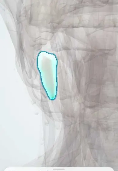
Digestive system · Head and neck
Lower right first premolar !
The lower right first premolar (tooth 44) is the tooth just behind the canine; it helps crush and grind food.
Opens in the Durgical app, no sign-up needed.
What it is
A transition tooth between the canine and the molars, with two cusps (cheek side and tongue side).
What it does
Crushes and tears food.
Key facts
- Joints with
- Alveolar socket of the mandible (gomphosis held by the periodontal ligament); bites against the opposing teeth
- Landmarks
- FDI 44, lower right first premolar: 1 root; two cusps; often removed for orthodontic space; erupts at about 10–12 years; nerve: inferior alveolar nerve (V3)
Relations
- In front
- The lower right canine
- Behind
- The lower right second premolar
- Above
- Meets the upper right canine and the upper right first premolar
- Medial
- Tongue and floor of the mouth; its root tip lies above the mylohyoid line
How to find it
Fourth from the midline on the lower right, behind the canine; the mental nerve leaves the bone below the area between it and the second premolar.
On imaging
A panoramic film shows the mental foramen as a round dark spot below the premolar roots, useful to mark before implants or flaps; bitewings show decay at its contacts.
Common problems
- Often the tooth removed to make space in orthodontic treatment.
Clinical notes
- Supplied by the inferior alveolar nerve (V3); an inferior alveolar nerve block or a mental (incisive) block numbs it
- Infection from its apex, above the attachment of the mylohyoid, spreads into the sublingual space and raises the floor of the mouth on that side
Anatomical variants
- A small extra cusp-like tubercle in the middle of its biting surface (dens evaginatus) occurs mainly on lower premolars and can fracture and open the pulp
Development
Replaces the primary lower right first molar (FDI 84). The primary molars are wider than their successors, and in the lower arch this spare room (leeway space) is larger than in the upper.
Related conditions
Test yourself
Think of the answer, then tap the question to see it.
Where does the mental foramen usually lie in relation to the lower right first premolar?
Below the space between the two lower premolars or below the second premolar.
Where does infection from the apex of the lower right first premolar spread?
Into the sublingual space, because the apex lies above the mylohyoid attachment; the floor of the mouth rises on that side.
Which primary tooth does the lower right first premolar replace?
The primary lower right first molar (FDI 84).
On a panoramic film, where does the mental foramen appear in relation to the lower right first premolar?
As a round dark spot below the premolar roots, usually between the two premolars or under the second.
Educational information, not medical advice. For any symptom, see a health professional.
Related
Sources: Anatomy, Head and Neck, Teeth; Anatomy, Permanent Dentition; Anatomy, Head and Neck, Tooth Eruption; Anatomy, Head and Neck: Alveolar Nerve; 23.3 The Mouth, Pharynx, and Esophagus (OpenStax (Rice University), 2022) · adapted, CC BY 4.0.
Spotted a mistake?We are always improving for the community. Tell us what is wrong and our team will review it.Report a mistake
Representative model, for educational purposes only.