Muscular system · Head and neck
Mylohyoid !
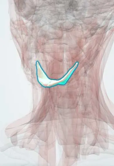
The mylohyoid is a sheet of muscle that forms the floor of the mouth; it lifts the floor of the mouth and the tongue when swallowing.
Official name: Mylohyoid muscle
Opens in the Durgical app, no sign-up needed.
What it is
A sheet of muscle forming the floor of the mouth.
What it does
Lifts the floor of the mouth and the tongue when swallowing.
Key facts
- Origin
- Mylohyoid line of the mandible
- Insertion
- Midline raphe and body of the hyoid
- Action
- Raises the floor of the mouth and the hyoid; depresses the mandible
- Nerve
- Nerve to mylohyoid (V3)
- Blood supply
- Submental artery, mylohyoid branch of the inferior alveolar artery
Relations
- Above
- Geniohyoid, genioglossus and hyoglossus; the sublingual gland, the submandibular duct, the lingual nerve and the hypoglossal nerve lie on its oral surface
- Below
- Anterior belly of the digastric, the superficial part of the submandibular gland, the facial artery and vein, and the submental lymph nodes
- Behind
- Free posterior border: the submandibular gland hooks round it, with its deep part and duct above the muscle, and the hypoglossal nerve passes deep to it into the floor of the mouth
- Medial
- Joins its partner at a midline raphe running from the chin to the hyoid bone
How to find it
It is not felt as a separate muscle: pressing up under the chin while the person swallows gives the firm floor of the mouth. In surgery it appears once the anterior belly of the digastric is retracted; pulling its posterior border forward exposes the lingual nerve, the submandibular duct and the hypoglossal nerve.
Examination
- Bimanual palpation of the floor of the mouth, one gloved finger inside and the other hand under the jaw, with the muscle between them: used to feel a stone in the submandibular duct, an enlarged gland or a mass in the sublingual or submandibular space
On imaging
Coronal CT or MRI shows it as a sling from the mylohyoid line of each side to the hyoid: the sublingual space lies above it, the submandibular space below. Contrast CT is chosen in Ludwig angina or a dental abscess to map spread around its free posterior border and to find gas or a drainable collection. A plunging ranula passes from the sublingual space through or behind it.
Clinical notes
- In Ludwig angina the swelling is woody and the floor of the mouth rises; airway control comes first, before draining the spaces
- Gaps in the muscle let sublingual gland tissue or a ranula pass into the neck (plunging ranula)
- In submandibular gland excision it is retracted forward to reach the deep part of the gland, the duct and the nerves above it
Anatomical variants
- Defects (hiatuses) in its front part are common; sublingual gland tissue or fat may bulge through them and mimic a mass under the jaw
Development
From the first pharyngeal arch, like the anterior belly of the digastric, which is why both receive the nerve to mylohyoid.
Related conditions
Test yourself
Think of the answer, then tap the question to see it.
Which two spaces does the mylohyoid separate, and where do they communicate?
The sublingual space above from the submandibular space below; they communicate around its free posterior border.
Why do infections of the lower second and third molars spread to the submandibular space?
Their roots lie below the mylohyoid line, which slopes down toward the front; premolar infections spread above it.
What is Ludwig angina and what is the first priority in its management?
A fast-spreading cellulitis of both submandibular and sublingual spaces, usually from a lower molar; airway control comes first, before drainage.
Which nerve supplies the mylohyoid?
The nerve to mylohyoid (V3), as the muscle comes from the first pharyngeal arch, like the anterior belly of the digastric.
Educational information, not medical advice. For any symptom, see a health professional.
Its names
- Official Latin (TA2)
- musculus mylohyoideus TA2 2165
- Official Spanish (DeCS)
- Músculo milohioideo
- English (TA2)
- Mylohyoid muscle
Related
Sources: Terminologia Anatomica (TA2); Anatomy, Head and Neck, Mylohyoid Muscle; Anatomy, Head and Neck: Suprahyoid Muscle.
Spotted a mistake?We are always improving for the community. Tell us what is wrong and our team will review it.Report a mistake
Representative model, for educational purposes only.