Joints and ligaments · Head and neck
Thyrohyoid membrane !
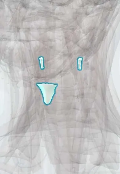
The thyrohyoid membrane is a sheet joining the thyroid cartilage to the hyoid bone in the upper neck, from which the larynx hangs.
Official name: Median thyrohyoid ligament
Opens in the Durgical app, no sign-up needed.
What it is
A sheet joining the thyroid cartilage to the hyoid.
What it does
Hangs the larynx from the hyoid.
Key facts
- Attaches
- Upper border of the thyroid cartilage to the hyoid
- Function
- Suspends the larynx; pierced by the internal laryngeal nerve and superior laryngeal artery
Relations
- Above
- Hyoid bone, attached to the upper edge of the back of its body, with a bursa between the membrane and the hyoid body
- Superficial
- Thyrohyoid, sternohyoid and the superior belly of the omohyoid
- Deep
- Pre-epiglottic space and the epiglottis; further out, the pyriform recesses
- Below
- Upper border of the thyroid cartilage and its superior horns
How to find it
The soft gap between the hyoid bone and the upper border of the thyroid cartilage is felt above the thyroid notch; it rises with the larynx on swallowing.
On imaging
CT shows a laryngocele as an air- or fluid-filled sac in the paraglottic space; an external one passes through the thyrohyoid membrane into the neck. A calcified cartilago triticea can look like a foreign body or calcified plaque on a radiograph.
Clinical notes
- A laryngocele bulges in the neck on coughing or straining, classically in wind instrument players and glassblowers; it can become infected (laryngopyocele)
- Supraglottic cancer that reaches the pre-epiglottic space lies just behind the membrane
- In supraglottic laryngectomy the internal laryngeal nerve is kept where it pierces the membrane, because sensation in the remaining larynx helps protect against aspiration
Test yourself
Think of the answer, then tap the question to see it.
Which nerve pierces the thyrohyoid membrane, and why is this site useful?
The internal laryngeal nerve, below the greater horn of the hyoid; it can be blocked there to numb the larynx above the vocal folds for awake airway procedures.
What is an external laryngocele?
A dilated laryngeal saccule that pushes out through the thyrohyoid membrane where the nerve and vessels pierce it; it bulges on coughing or straining.
What do the thickened parts of the thyrohyoid membrane form?
The median thyrohyoid ligament in the midline and the lateral thyrohyoid ligaments at the back edges, which often hold a small cartilago triticea.
Educational information, not medical advice. For any symptom, see a health professional.
Its names
- Official Latin (TA2)
- ligamentum thyreohyoideum medianum TA2 1652
- Official Spanish (DeCS)
- Membrana tirohioidea
- English (TA2)
- Median thyrohyoid ligament
- Also called
- ligamentum hyothyreoideum medium
Sources: Terminologia Anatomica (TA2); Anatomy, Head and Neck, Thyrohyoid Membrane.
Spotted a mistake?We are always improving for the community. Tell us what is wrong and our team will review it.Report a mistake
Representative model, for educational purposes only.