Muscular system · Head and neck
Thyrohyoid !
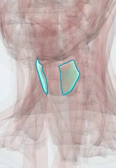
The thyrohyoid is a short strap muscle from the thyroid cartilage to the hyoid bone; it brings the larynx and the hyoid together.
Official name: Thyrohyoid muscle
Opens in the Durgical app, no sign-up needed.
What it is
A short strap muscle from the thyroid cartilage to the hyoid.
What it does
Brings the larynx and hyoid together.
Key facts
- Origin
- Oblique line of the thyroid cartilage
- Insertion
- Greater horn and body of the hyoid
- Action
- Depresses the hyoid or raises the larynx
- Nerve
- C1 via the hypoglossal nerve
- Blood supply
- Superior thyroid artery
Relations
- Deep
- Thyrohyoid membrane, with the internal laryngeal nerve and the superior laryngeal artery passing through it
- Superficial
- Sternohyoid and the superior belly of the omohyoid
- Below
- Continuous with the sternothyroid at the oblique line of the thyroid cartilage
How to find it
It fills the gap between the thyroid cartilage and the hyoid bone at the side of the midline; it is felt as soft tissue over the thyrohyoid membrane that rises with the larynx on swallowing.
Examination
- Laryngeal elevation on palpation: with fingers on the hyoid bone and the thyroid cartilage during a dry swallow, the cartilage should rise and close up to the hyoid; a reduced rise suggests weak laryngeal elevation and a risk of aspiration
On imaging
On ultrasound and CT it lies against the thyrohyoid membrane, lateral to the midline, between the hyoid and the thyroid cartilage. It is a landmark for an ultrasound-guided superior laryngeal nerve block, where the internal branch pierces the membrane below the greater horn of the hyoid. An infrahyoid thyroglossal duct cyst is often embedded in the strap muscles here; ultrasound is the first test.
Clinical notes
- The internal laryngeal nerve is blocked near the greater horn of the hyoid, where it pierces the thyrohyoid membrane deep to this muscle, to numb the larynx for awake airway procedures
- A thyroglossal duct cyst sits in the midline at or just below the hyoid bone, deep to the strap muscles, and moves up when the person swallows or sticks out the tongue
- In hyoid suspension surgery for obstructive sleep apnoea the hyoid bone is fixed forward and down onto the thyroid cartilage, across this muscle, to widen the airway behind the tongue base
Development
Shares the strap-muscle premuscle mass with the tongue muscles, entered early by the hypoglossal and upper cervical nerves, which fits its C1 supply carried by the hypoglossal nerve.
Test yourself
Think of the answer, then tap the question to see it.
Which strap muscle is not supplied by the ansa cervicalis, and what supplies it?
The thyrohyoid: its C1 fibres leave the hypoglossal nerve as the nerve to thyrohyoid.
Which nerve and artery pierce the thyrohyoid membrane deep to the thyrohyoid?
The internal branch of the superior laryngeal nerve and the superior laryngeal artery.
Where is the internal laryngeal nerve blocked to numb the larynx for awake airway procedures?
Near the greater horn of the hyoid, where it pierces the thyrohyoid membrane deep to the thyrohyoid.
Educational information, not medical advice. For any symptom, see a health professional.
Its names
- Official Latin (TA2)
- musculus thyreohyoideus TA2 2174
- Official Spanish (DeCS)
- Músculo tirohioideo
- English (TA2)
- Thyrohyoid muscle
Related
Sources: Terminologia Anatomica (TA2); Anatomy, Head and Neck, Thyroid Muscles; Anatomy, Head and Neck: Anterior Cervical Region; Anatomy of the Human Body (20th ed.): Development of the Muscles (Lea & Febiger (Gray's Anatomy, 1918), 1918) · adapted, Public domain; 11.3 Axial Muscles of the Head, Neck, and Back (OpenStax (Rice University), 2022) · adapted, CC BY 4.0.
Spotted a mistake?We are always improving for the community. Tell us what is wrong and our team will review it.Report a mistake
Representative model, for educational purposes only.