Skeletal system · Head and neck
Hyoid bone !
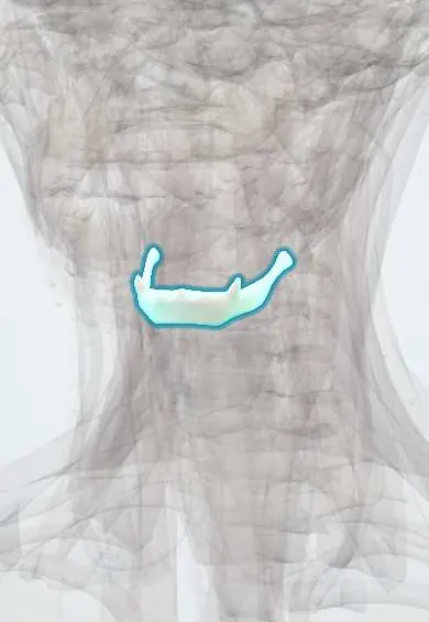
The hyoid is a U-shaped bone in the upper neck that touches no other bone; it anchors the tongue and the muscles that move the larynx when swallowing.
Opens in the Durgical app, no sign-up needed.
What it is
A U-shaped bone in the upper neck that touches no other bone.
What it does
Anchors the tongue and the muscles that move the larynx when swallowing.
Key facts
- Joints with
- None (floats; held by muscles and ligaments)
- Landmarks
- Body, greater and lesser horns; level C3
Relations
- Above
- Floor of the mouth and the tongue: mylohyoid, geniohyoid, hyoglossus and the middle pharyngeal constrictor attach here
- Below
- Thyroid cartilage, joined by the thyrohyoid membrane (pierced by the internal laryngeal nerve and superior laryngeal vessels); sternohyoid, omohyoid and thyrohyoid attach below
- Behind
- Epiglottis, linked by the hyoepiglottic ligament, with the pre-epiglottic space between them; the pharynx behind
- Lateral
- The greater horns reach toward the carotid bifurcation; the hypoglossal nerve and lingual artery run just above them
- Superficial
- Platysma and skin in the angle between the chin and the neck
How to find it
With the neck slightly extended it is felt in the angle between the floor of the mouth and the front of the neck, above the thyroid cartilage; it can be moved side to side and rises on swallowing.
On imaging
CT shows fractures of the horns and body; in trauma and forensic imaging an unfused joint between the body and a greater horn can be mistaken for a fracture.
Clinical notes
- Thyroglossal cyst surgery removes the middle part of the hyoid body with the cyst and tract (Sistrunk operation), which lowers recurrence
- Hyoid suspension is one of the surgical options for obstructive sleep apnoea
- The greater horn can be felt and mistaken for a lump in a thin neck
Anatomical variants
- The stylohyoid ligament, from the styloid process to the lesser horn, may be partly ossified or the styloid process elongated; a few of these people have throat or neck pain on swallowing or turning the head (Eagle syndrome)
Development
Develops from the second (lesser horns, upper body) and third (greater horns, lower body) pharyngeal arch cartilages; the greater horns fuse to the body in adult life or never.
Related conditions
Test yourself
Think of the answer, then tap the question to see it.
What does a fractured greater horn of the hyoid bone suggest at autopsy?
Neck compression, more with manual strangulation than hanging; but it is often intact, especially in young people whose hyoid has not fused.
Why does a thyroglossal cyst rise on tongue protrusion, and what does its surgery remove?
The thyroglossal duct passes by or through the hyoid body; the Sistrunk operation removes the middle of the hyoid body with the cyst and tract.
Which muscle groups attach to the hyoid bone, and what do they do in swallowing?
The suprahyoid (digastric, stylohyoid, mylohyoid, geniohyoid) and infrahyoid muscles; they raise and lower the larynx.
Which nerve pierces the thyrohyoid membrane between the hyoid bone and the thyroid cartilage?
The internal laryngeal nerve, with the superior laryngeal vessels.
Educational information, not medical advice. For any symptom, see a health professional.
Its names
- Official Latin (TA2)
- os hyoideum TA2 876
- Official Spanish (DeCS)
- Hueso Hioides DeCS D006928
- English (TA2)
- Hyoid bone
- Also called
- os hyoides · os linguae · lingual bone
Sources: Terminologia Anatomica (TA2); DeCS; Anatomy, Head and Neck: Hyoid Bone.
Spotted a mistake?We are always improving for the community. Tell us what is wrong and our team will review it.Report a mistake
Representative model, for educational purposes only.