Digestive system · Head and neck
Upper left canine !
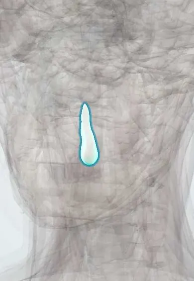
The upper left canine (tooth 23) is the pointed eye tooth at the corner of the mouth; it has the longest root and grips and tears food.
Opens in the Durgical app, no sign-up needed.
What it is
The corner tooth: one pointed cusp and the longest root of all the teeth.
What it does
Grips and tears food, and guides the jaw when it slides sideways.
Key facts
- Joints with
- Alveolar socket of the maxilla (gomphosis held by the periodontal ligament); bites against the opposing teeth
- Landmarks
- FDI 23, upper left canine: 1 root, the longest of all teeth; single pointed cusp; the 'eye tooth', often impacted in the palate; erupts at about 11–12 years; nerve: anterior superior alveolar nerve (V2)
Relations
- Medial
- The upper left lateral incisor
- Lateral
- The upper left first premolar
- In front
- Corner of the upper lip, with the canine eminence under the mucosa
- Below
- Meets the lower left canine and the lower left first premolar
How to find it
Third from the midline on the upper left, the pointed tooth at the bend of the arch; the root's ridge, the canine eminence, can be felt under the lip running toward the nose.
On imaging
Panoramic radiographs show an impacted canine; cone-beam CT shows whether it lies on the palatal or the labial side and whether it is resorbing the incisor roots.
Clinical notes
- Supplied by the anterior superior alveolar nerve (V2); the long root means the anaesthetic must reach higher in the vestibule than for the incisors
- An impacted canine can resorb the roots of the neighbouring lateral and central incisors without symptoms
- Its long, strong root makes it a valued support for bridges
Development
Replaces the primary upper left canine (FDI 63). Its crown starts forming high in the maxilla, beside the nose, which explains its long and late path into the mouth.
Memory aid
SLOB (Same Lingual, Opposite Buccal): between two films with the tube shifted, an impacted canine that follows the tube is palatal, one that goes the other way is buccal.
Test yourself
Think of the answer, then tap the question to see it.
Which imaging shows whether an impacted upper left canine lies palatal or labial?
Cone-beam CT, which also shows whether it is resorbing the incisor roots.
What silent complication can an impacted upper left canine cause?
Resorption of the roots of the neighbouring lateral and central incisors.
Why must anaesthetic for the upper left canine reach higher in the vestibule than for the incisors?
Its root is the longest of all the teeth.
Why is the upper left canine a good support for a bridge?
It has the longest root of any tooth, which anchors it firmly.
Educational information, not medical advice. For any symptom, see a health professional.
Related
Sources: Anatomy, Head and Neck, Teeth; Anatomy, Permanent Dentition; Anatomy, Head and Neck, Tooth Eruption; Anatomy, Head and Neck: Alveolar Nerve; 23.3 The Mouth, Pharynx, and Esophagus (OpenStax (Rice University), 2022) · adapted, CC BY 4.0.
Spotted a mistake?We are always improving for the community. Tell us what is wrong and our team will review it.Report a mistake
Representative model, for educational purposes only.