Digestive system · Head and neck
Upper left lateral incisor !
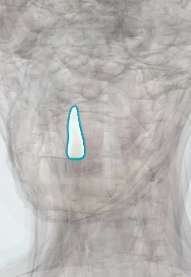
The upper left lateral incisor (tooth 22) is the flat front tooth next to the central incisor; it helps cut food.
Opens in the Durgical app, no sign-up needed.
What it is
A front tooth beside the central incisor, a little smaller, with a cutting edge and one root.
What it does
Bites into and cuts food.
Key facts
- Joints with
- Alveolar socket of the maxilla (gomphosis held by the periodontal ligament); bites against the opposing teeth
- Landmarks
- FDI 22, upper left lateral incisor: 1 root; smaller than the central incisor; often small, peg-shaped or missing; erupts at about 8–9 years; nerve: anterior superior alveolar nerve (V2)
Relations
- Medial
- The upper left central incisor
- Lateral
- The upper left canine
- In front
- Upper lip and the orbicularis oris
- Below
- Its palatal surface meets the lower left lateral incisor and the lower left canine
How to find it
Second from the midline on the upper left, between the left central incisor and the canine; smaller than the central, which helps tell it apart.
On imaging
A panoramic radiograph shows whether it is present and how close the left canine's crown sits to its root; a periapical reveals an enamel fold (dens invaginatus) inside the crown.
Common problems
- Sometimes small or missing from birth (more often the upper one).
Clinical notes
- Supplied by the anterior superior alveolar nerve (V2), a branch of the infraorbital nerve; local infiltration over the apex is enough because the labial bone is thin
- In cleft lip and palate the cleft runs between the lateral incisor and the canine; one-sided clefts are more often on the left, and on the cleft side this tooth is often missing, doubled or malformed
Anatomical variants
- Missing from birth more often than most teeth, or reduced to a small conical (peg) crown
Development
Replaces the primary upper left lateral incisor (FDI 62). It forms where the premaxilla meets the rest of the maxilla, which explains its link with clefts.
Related conditions
Test yourself
Think of the answer, then tap the question to see it.
Which developmental anomaly most often affects the upper left lateral incisor?
Dens invaginatus: an enamel fold that dips toward the pulp.
Between which teeth does a cleft of the lip and palate run?
Between the lateral incisor and the canine; on the cleft side the lateral incisor is often missing, doubled or malformed.
Which nerve supplies the upper left lateral incisor?
The anterior superior alveolar nerve (V2), a branch of the infraorbital nerve; infiltration over the apex suffices because the labial bone is thin.
Which radiograph shows whether the upper left lateral incisor is present and how close the canine lies to its root?
A panoramic radiograph.
Educational information, not medical advice. For any symptom, see a health professional.
Related
Sources: Anatomy, Head and Neck, Teeth; Anatomy, Permanent Dentition; Anatomy, Head and Neck, Tooth Eruption; Anatomy, Head and Neck: Alveolar Nerve; 23.3 The Mouth, Pharynx, and Esophagus (OpenStax (Rice University), 2022) · adapted, CC BY 4.0.
Spotted a mistake?We are always improving for the community. Tell us what is wrong and our team will review it.Report a mistake
Representative model, for educational purposes only.