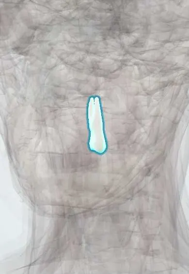
Digestive system · Head and neck
Upper left first premolar !
The upper left first premolar (tooth 24) is the tooth just behind the canine; it helps crush and grind food.
Opens in the Durgical app, no sign-up needed.
What it is
A transition tooth between the canine and the molars, with two cusps (cheek side and tongue side); the upper first premolar often has two roots.
What it does
Crushes and tears food.
Key facts
- Joints with
- Alveolar socket of the maxilla (gomphosis held by the periodontal ligament); bites against the opposing teeth
- Landmarks
- FDI 24, upper left first premolar: usually 2 roots (buccal and palatal); two cusps; often removed for orthodontic space; erupts at about 10–11 years; nerve: middle superior alveolar nerve (V2)
Relations
- In front
- The upper left canine
- Behind
- The upper left second premolar
- Lateral
- Cheek and buccinator across the buccal vestibule
- Below
- Meets the lower left first premolar and the lower left second premolar
How to find it
Behind the upper left canine, the fourth tooth from the midline; seen from the cheek side its tall buccal cusp looks like a small canine.
On imaging
Bitewing radiographs catch decay hidden at its contacts with the canine and the second premolar; an angled periapical splits the two roots so neither canal is missed.
Common problems
- Often the tooth removed to make space in orthodontic treatment.
Clinical notes
- Supplied by the middle superior alveolar nerve, an inconstant branch; when it is absent, fibres from the anterior and posterior superior alveolar nerves take over, and buccal infiltration still numbs the tooth
- When the maxillary sinus extends far forward its floor can reach above these roots, so the ache of sinusitis may be felt in this tooth
Anatomical variants
- Many have a single root holding 2 canals instead of 2 separate roots; rarely there are 3 roots (two buccal, one palatal)
Development
Replaces the primary upper left first molar (FDI 64). The primary molars are wider than the premolars that follow them, and the spare room (leeway space) helps the permanent teeth line up.
Related conditions
Test yourself
Think of the answer, then tap the question to see it.
Which premolar usually has 2 root canals?
The upper first premolar, such as the upper left first premolar; a missed canal is a classic cause of failed root canal treatment.
Why may the ache of sinusitis be felt in the upper left first premolar?
When the maxillary sinus extends far forward, its floor can reach above this tooth's roots.
What is leeway space?
The spare room left because the primary molars are wider than the premolars that replace them, such as the upper left first premolar; it helps the permanent teeth line up.
Which radiograph best shows hidden decay between the upper left first premolar and its neighbours?
The bitewing radiograph.
Educational information, not medical advice. For any symptom, see a health professional.
Related
Sources: Anatomy, Head and Neck, Teeth; Anatomy, Permanent Dentition; Anatomy, Head and Neck, Tooth Eruption; Anatomy, Head and Neck: Alveolar Nerve; 23.3 The Mouth, Pharynx, and Esophagus (OpenStax (Rice University), 2022) · adapted, CC BY 4.0.
Spotted a mistake?We are always improving for the community. Tell us what is wrong and our team will review it.Report a mistake
Representative model, for educational purposes only.