
Vogt-Koyanagi-Harada disease
An autoimmune attack on pigment cells causing bilateral uveitis with retinal detachments, headache and hearing loss. It is more common in Latin Americans.
What it is
A multisystem autoimmune disease directed against melanocytes, affecting the eyes, meninges, inner ear and skin. In the eye it causes a bilateral granulomatous panuveitis with multiple serous retinal detachments and optic disc swelling. Without early, strong immunosuppression it becomes chronic and recurrent and leaves a depigmented 'sunset glow' fundus.
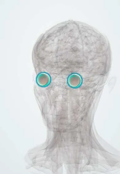
See it in 3D
Who gets it
More frequent in people with pigmented skin, including Latin American, Asian and Middle Eastern populations; usually adults aged 20 to 50, more often women. It is one of the main causes of non-infectious uveitis in Colombia and Latin America.
Causes
- Autoimmunity against melanocyte proteins in a genetically predisposed person (HLA-DR4 and related types)
- Possible viral trigger
Risk factors
- Pigmented skin
- Female sex
- Certain HLA types
Pathophysiology
T cells target melanocytes in the choroid, meninges, inner ear and skin. Choroidal inflammation thickens the choroid and leaks fluid under the retina; later, loss of melanocytes depigments the fundus, skin and hair.
Classification
- Prodromal (flu-like, headache, neck stiffness, tinnitus)
- Acute uveitic (bilateral blurred vision, serous detachments)
- Convalescent (depigmentation, vitiligo, poliosis)
- Chronic recurrent
Symptoms and signs
- Headache, neck stiffness and malaise
- Ringing in the ears and hearing loss
- Sudden blurred vision in both eyes
- Later: white patches of skin, whitened eyelashes and hair loss
Diagnosis
- Clinical
- Bilateral panuveitis with multiple serous retinal detachments and disc swelling, without previous eye trauma or surgery.
- Tests
- Lumbar puncture may show a lymphocytic pleocytosis; syphilis, tuberculosis and sarcoidosis are excluded.
- Imaging
- OCT shows subretinal fluid and a thick choroid; fluorescein angiography shows multiple pinpoint leaks; ultrasound shows choroidal thickening.
Differential diagnosis
- Sympathetic ophthalmia (previous eye injury or surgery)
- Central serous chorioretinopathy
- Posterior scleritis
- Syphilis, tuberculosis, sarcoidosis
- Uveal lymphoma
Goal of treatment
Suppress inflammation early and completely to prevent chronic disease and vision loss.
Treatment
- Medical
- High-dose systemic corticosteroids (often intravenous at first) tapered slowly over many months.
- Medical
- Early immunosuppressive or biologic drugs to spare steroids and prevent recurrences.
- Follow-up
- Regular ophthalmology and OCT monitoring for recurrence, cataract and glaucoma.
Complications
- Cataract
- Glaucoma
- Choroidal neovascularisation
- Subretinal fibrosis
- Permanent depigmentation and hearing loss
Prognosis
Good when treated early and adequately; chronic recurrent disease risks permanent visual loss.
Test yourself
Think of the answer, then tap the question to see it.
Which tissues does Vogt-Koyanagi-Harada disease affect?
Melanocyte-containing tissues: the eye (uvea), meninges, inner ear and skin.
What is the typical eye finding?
Bilateral panuveitis with multiple serous retinal detachments.
What history separates it from sympathetic ophthalmia?
No previous penetrating eye injury or eye surgery.
Connections
Educational information, not medical advice. For any symptom, see a health professional.
Sources: Vogt-Koyanagi-Harada Syndrome (StatPearls Publishing (NCBI Bookshelf), 2026).
Spotted a mistake?We are always improving for the community. Tell us what is wrong and our team will review it.Report a mistake
Representative model, for educational purposes only.