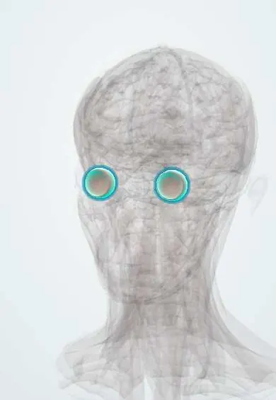
Choroid !
The choroid is a layer rich in blood vessels between the retina and the white of the eye; it feeds the outer retina.
Opens in the Durgical app, no sign-up needed.
What it is
A layer full of blood vessels between the retina and the sclera.
What it does
Feeds the outer retina; inflamed in posterior uveitis.
Key facts
- Where
- Between the retina and the sclera (posterior uvea)
- Blood supply
- Short posterior ciliary arteries
- Function
- Nourishes the outer retina (photoreceptors)
Relations
- In front
- Continuous with the ciliary body at the ora serrata
- Behind
- Ends at the optic disc, where the optic nerve leaves the eye
- Superficial
- Sclera, across the potential suprachoroidal space, where fluid or blood can collect (choroidal detachment)
- Deep
- Bruch membrane, the retinal pigment epithelium and the outer retina (photoreceptors), which it nourishes
- Contents
- From outside in: layers of large (Haller) and medium (Sattler) vessels, the choriocapillaris and Bruch membrane; blood leaves through the vortex veins, usually four, one per quadrant
How to find it
On fundoscopy it gives the orange-red background of the fundus; in thin or myopic fundi its large vessels show through as a striped (tigroid) pattern. Its full thickness is seen only on OCT or ultrasound.
On imaging
Indocyanine green angiography shows the choroidal circulation best; enhanced-depth OCT measures its thickness; B-scan ultrasound shows a melanoma as a dome- or mushroom-shaped mass with low internal reflectivity.
Clinical notes
- Posterior uveitis (choroiditis) from toxoplasmosis, tuberculosis or syphilis; toxoplasma retinochoroiditis is a frequent infectious cause, with a white active focus beside an old pigmented scar
- Choroidal tubercles on fundoscopy support the diagnosis of miliary tuberculosis
- Choroidal rupture after blunt trauma appears as a curved yellow-white streak concentric to the optic disc
- Choroidal detachment can follow eye surgery when the eye pressure falls too low
Development
Forms from neural crest mesenchyme around the optic cup; incomplete closure of the optic (choroidal) fissure leaves a coloboma, typically inferonasal.
Related conditions
Test yourself
Think of the answer, then tap the question to see it.
What is the blood supply of the fovea?
Only the choriocapillaris of the choroid: the fovea has no retinal vessels.
Which layer of the choroid is involved in age-related macular degeneration, and how?
Bruch membrane: drusen build up there, and in the wet form new vessels break through it.
What is the most common primary intraocular cancer in adults, and what is its common benign look-alike?
Choroidal (uveal) melanoma; a flat choroidal naevus is the common benign look-alike.
What do choroidal tubercles on fundoscopy support?
The diagnosis of miliary tuberculosis.
Educational information, not medical advice. For any symptom, see a health professional.
Its names
- Official Latin (TA2)
- chorioidea TA2 6774
- Official Spanish (DeCS)
- Coroides
- English (TA2)
- Choroid
- Also called
- chorioides
Related
Sources: Terminologia Anatomica (TA2); Uveitis; Anatomy, Head and Neck, Eye.
Spotted a mistake?We are always improving for the community. Tell us what is wrong and our team will review it.Report a mistake
Representative model, for educational purposes only.