Nervous system · Head and neck
Optic nerve !
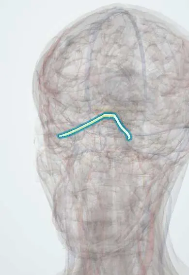
The optic nerve is the cable of about a million nerve fibres carrying vision from the eye to the brain; glaucoma damages it.
Opens in the Durgical app, no sign-up needed.
What it is
The cable of about a million nerve fibres from the eye to the brain.
What it does
Carries vision; damaged by glaucoma and inflamed in optic neuritis.
Key facts
- Roots
- Retinal ganglion cells -> lateral geniculate body; leaves the orbit through the optic canal
- Moves
- None
- Feeling from
- Vision
Relations
- Course
- From the optic disc back and medially through the orbit inside the cone of the rectus muscles, through the common tendinous ring and the optic canal of the sphenoid bone, to the optic chiasm above the sella turcica
- Contents
- Central retinal artery and vein, which enter it a short distance behind the eyeball and reach the retina at the disc
- Below
- Ophthalmic artery, below it in the optic canal before crossing to its medial side in the orbit
- Medial
- Sphenoid sinus and posterior ethmoid cells, separated by thin bone, a risk in sinus surgery
- Lateral
- Near the apex, the ciliary ganglion between it and the lateral rectus, with the short ciliary nerves running forward around it; the ophthalmic artery and nasociliary nerve cross above it from lateral to medial
How to find it
The optic disc is seen with the ophthalmoscope: colour, sharpness of the margins and the size of the central cup (cup-to-disc ratio) are recorded. The orbital part is followed on MRI from the back of the eyeball to the optic canal.
Examination
- Swinging flashlight test: when the light moves to the affected eye both pupils dilate instead of constricting (RAPD)
- Visual acuity and colour vision (colour plates, red desaturation): both fall early in optic nerve disease
- Confrontation and formal visual fields: central scotoma in optic neuritis, enlarged blind spot in papilloedema
- Fundoscopy: swollen disc (papilloedema, papillitis), pale disc (optic atrophy) or a large cup (glaucoma)
On imaging
Contrast MRI of the orbits with fat suppression shows an enhancing nerve in optic neuritis, and brain MRI shows white matter lesions that predict multiple sclerosis; optical coherence tomography measures the thickness of the nerve fibre layer around the disc.
Common problems
- Glaucoma slowly destroys its fibres.
- Optic neuritis: painful blurring of one eye, sometimes the first sign of multiple sclerosis.
- Compression by tumours or swelling.
How it is diagnosed
Visual acuity, colour vision, pupil test, visual fields, OCT and MRI.
Treatment
- Lower eye pressure for glaucoma.
- Steroids in some optic neuritis.
- Treat the cause of compression.
Clinical notes
- Papilloedema is bilateral disc swelling from raised intracranial pressure; vision is usually preserved early, apart from an enlarged blind spot, unlike optic neuritis
- Anterior ischaemic optic neuropathy: sudden painless loss of vision with a swollen disc; in older people giant cell arteritis is excluded urgently
- Fractures through the optic canal can injure the nerve (traumatic optic neuropathy)
- A cut nerve does not regenerate: loss of vision from optic nerve damage is usually permanent
Anatomical variants
- Disc and cup size vary widely: a large disc normally has a large cup, so a big cup that is the same in both eyes may be physiological, while a difference between the eyes raises concern for glaucoma
- Tilted disc: an oval, obliquely set disc, common in myopia; it can give upper temporal field defects that mimic a chiasmal lesion but cross the vertical midline
Development
Grows from the optic stalk, an outpouching of the forebrain (diencephalon), which is why it is central nervous tissue with meninges.
Related conditions
- Leber hereditary optic neuropathy (LHON)
- Methanol poisoning
- Open-angle glaucoma
- Multiple sclerosis
- Giant cell arteritis
- Idiopathic intracranial hypertension
- Neurofibromatosis (types 1 and 2)
- Neuromyelitis optica spectrum disorder
- Optic neuritis
- Thyroid eye disease (Graves orbitopathy)
Test yourself
Think of the answer, then tap the question to see it.
Why does raised intracranial pressure swell the optic disc?
The optic nerve is wrapped in meninges with a subarachnoid space continuous with the brain's, so the pressure reaches the disc (papilloedema).
What are the classic features of optic neuritis?
Pain on eye movement, reduced acuity and colour vision and a relative afferent pupillary defect; the disc often looks normal. It can be an early sign of multiple sclerosis.
What does the swinging flashlight test show with a damaged optic nerve?
A relative afferent pupillary defect: when the light moves to the affected eye, both pupils dilate instead of constricting.
Through which opening does the optic nerve leave the orbit, and where does it end?
Through the optic canal of the sphenoid bone, to the optic chiasm above the sella turcica.
Why is the optic nerve considered a tract of the brain?
It grows from the optic stalk of the forebrain, is myelinated by oligodendrocytes and is wrapped in meninges.
Educational information, not medical advice. For any symptom, see a health professional.
Its names
- Official Latin (TA2)
- nervus opticus TA2 6183
- Official Spanish (DeCS)
- Nervio óptico
- English (TA2)
- Optic nerve
- Also called
- cranial nerve II · fasciculus opticus
Related
Sources: Terminologia Anatomica (TA2); Neuroanatomy, Cranial Nerve 2 (Optic); Anatomía humana (4.ª ed.), tomo 1, cap. 45: Estructuras accesorias del ojo.
Spotted a mistake?We are always improving for the community. Tell us what is wrong and our team will review it.Report a mistake
Representative model, for educational purposes only.