Age-related macular degeneration !
Age-related macular degeneration is wear of the macula, the centre of the retina, in older adults; it blurs central vision while side vision remains.
Updated 2026-10-29
Structures involved
Tap one to see what it is and open it in 3D.
In histology
In physiology
Why it happens
- How organs age: normal changes vs disease
- Oxidative stress and free-radical injury
- Cellular aging: telomeres, DNA damage, senescence and proteostasis
What it is
Progressive degeneration of the macula in older adults, affecting the retinal pigment epithelium, Bruch membrane and choroid. It damages central vision while side vision is usually kept.
Who gets it
A leading cause of irreversible vision loss in older adults worldwide. It becomes more frequent as populations age, including in Latin America.
Causes
- Age-related changes of the retinal pigment epithelium and Bruch membrane
- Genetic susceptibility (complement pathway genes and others)
- Oxidative stress and chronic inflammation
Risk factors
- Age over about 55
- Smoking (the main modifiable factor)
- Family history
- White ancestry
- Cardiovascular disease, hypertension, dyslipidaemia
- Obesity and a diet low in antioxidants
Pathophysiology
Waste deposits called drusen build up between the retinal pigment epithelium and Bruch membrane, disturbing exchange with the choroid. In the dry (atrophic) form, epithelium and photoreceptors slowly die. In the wet (neovascular) form, new vessels grow from the choroid under the retina, leak fluid and bleed, causing faster loss of central vision and scarring.
Classification
- Early and intermediate: drusen and pigment changes
- Late dry: geographic atrophy
- Late wet: neovascular (exudative)
Symptoms and signs
- Gradual blurring of central vision; difficulty reading and recognising faces
- Straight lines look wavy (metamorphopsia)
- A dark or empty patch in the centre of vision
- Needs more light; reduced contrast
- Wet form: faster, sometimes sudden, central loss
Diagnosis
- Clinical
- Dilated fundus examination shows drusen, pigment changes, atrophy or haemorrhage and exudate. The Amsler grid detects distortion and is used for home monitoring.
- Tests
- Visual acuity testing. Fluorescein or indocyanine green angiography identifies and types choroidal neovascularisation.
- Imaging
- Optical coherence tomography (OCT) shows drusen, atrophy and fluid, and guides treatment; OCT angiography shows new vessels without dye. Fundus autofluorescence maps atrophy.
Differential diagnosis
- Diabetic macular oedema
- Central serous chorioretinopathy
- Macular hole or epiretinal membrane
- Hereditary macular dystrophies
- Myopic choroidal neovascularisation
- Cataract (when blurring is diffuse)
Goal of treatment
Slow progression, treat neovascular disease quickly to keep central vision, and help people make the most of their remaining sight.
Treatment
- Lifestyle
- Stopping smoking, a diet rich in green leafy vegetables and fish, and control of cardiovascular risk factors.
- Medical
- Antioxidant vitamin and mineral supplements of a specific formula slow progression in intermediate disease; they are not shown to prevent early disease.
- Medical
- Wet AMD: repeated intravitreal anti-VEGF injections, the standard of care, that dry the macula and stabilise or improve vision.
- Follow-up
- Home Amsler grid checks and regular reviews with OCT; injection intervals are adjusted to disease activity.
- Rehabilitation
- Low-vision rehabilitation: magnifiers, lighting, large print and training to use peripheral vision.
Complications
- Legal blindness in the centre of vision
- Subretinal haemorrhage and disciform scarring
- Visual hallucinations (Charles Bonnet syndrome)
- Falls, depression and loss of independence
Prognosis
Dry AMD usually progresses slowly over years. Anti-VEGF treatment has greatly improved the outlook in wet AMD, though long-term treatment is often needed and some vision may still be lost. Peripheral vision usually remains.
Prevention
- Not smoking
- Healthy diet and cardiovascular risk control
- Regular eye examinations from middle age, especially with a family history
Test yourself
Think of the answer, then tap the question to see it.
What are drusen?
Yellow extracellular deposits between the retinal pigment epithelium and Bruch membrane, the hallmark of early AMD.
What distinguishes wet from dry AMD?
Wet AMD has choroidal neovascularisation that leaks and bleeds; dry AMD shows drusen and slow atrophy without new vessels.
What is the standard treatment for neovascular AMD?
Repeated intravitreal anti-VEGF injections.
Which simple test detects metamorphopsia and is used at home?
The Amsler grid.
What is the main modifiable risk factor for AMD?
Smoking.
Related conditions
Educational information, not medical advice. For any symptom, see a health professional.
Sources: Macular Degeneration (StatPearls (NCBI Bookshelf), 2023).
Spotted a mistake?We are always improving for the community. Tell us what is wrong and our team will review it.Report a mistake
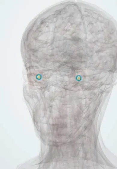
Representative model, for educational purposes only.