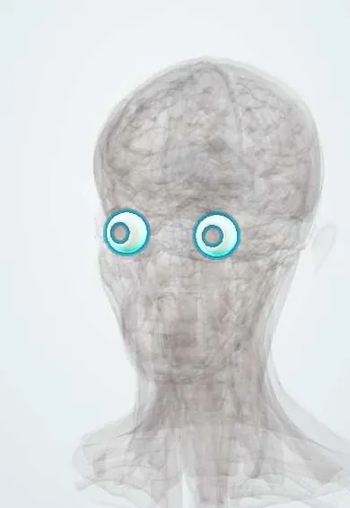
Sclera !
The sclera is the tough white outer coat of the eye; it protects the eyeball and anchors the muscles that move it.
Opens in the Durgical app, no sign-up needed.
What it is
The tough white outer coat of the eye.
What it does
Protects the eye and anchors the eye muscles.
Key facts
- Where
- Posterior five sixths of the outer coat
- Blood supply
- Episcleral and anterior ciliary vessels
- Nerve
- Ciliary nerves
- Function
- Protects the eye, anchors the eye muscles
Relations
- In front
- Cornea at the limbus, where Schlemm canal runs within it
- Behind
- Optic nerve, whose dural sheath blends with it
- Superficial
- Episclera and Tenon capsule (fascia bulbi), and at the front the conjunctiva
- Deep
- Choroid and ciliary body, across the suprachoroidal space
- Contents
- Canals for the posterior ciliary arteries and nerves around the optic nerve, the vortex veins behind the equator and the anterior ciliary vessels near the limbus
How to find it
The white of the eye, seen through the clear conjunctiva; the tendons of the extraocular muscles such as the superior rectus and lateral rectus insert on it in front of the equator. Its back part is seen only on imaging or at surgery.
Examination
- Phenylephrine drop test: superficial episcleral vessels blanch (episcleritis), deep scleral vessels do not (scleritis)
- Inspection in daylight: a bluish-violet hue suggests scleritis; tenderness to gentle pressure through the lid supports it
- Exophthalmometry measures how far the globe sits forward: high in thyroid eye disease or an orbital mass, low (enophthalmos) after a blow-out fracture of the orbital floor
On imaging
B-scan ultrasound shows posterior scleritis as a thickened coat with fluid in Tenon space around the optic nerve (T sign); CT helps when a hidden rupture is suspected. B-scan also looks inside the eye when a dense cataract or vitreous blood hides it. After trauma CT comes first; MRI waits until a metal foreign body is excluded.
Clinical notes
- Necrotizing scleritis can thin the sclera until the choroid shows; painless thinning in long-standing rheumatoid arthritis is scleromalacia perforans
- A scleral buckle sewn onto it indents the wall to close a retinal tear
- In high myopia the globe is too long for its optics (normal adult axial length is about 23–24 mm) and can bulge backward (posterior staphyloma); such eyes are also prone to retinal tears
- Open globe: a full-thickness wound of sclera or cornea is suggested by a peaked pupil, a shallow anterior chamber or dark uveal tissue at the wound
- Sympathetic ophthalmia: a rare inflammation of the uninjured eye weeks to months after a penetrating injury to the other
Development
Forms from neural crest mesenchyme that condenses around the optic cup.
Related conditions
- Ocular trauma: hyphema and open globe injury
- Scleritis and episcleritis
- Retinal detachment
- Conjunctivitis
Test yourself
Think of the answer, then tap the question to see it.
Where is the sclera thinnest, and where does it usually rupture after blunt trauma?
Just behind the insertions of the rectus muscles; this zone and the limbus are the usual rupture sites.
How does scleritis differ from episcleritis?
Scleritis gives deep, boring pain that can wake the patient and a violet-red tender eye, often with systemic disease; episcleritis is milder and usually self-limiting.
What does the phenylephrine drop test show in a red eye with suspected scleral inflammation?
Superficial episcleral vessels blanch (episcleritis); deep scleral vessels do not (scleritis).
What does a blue sclera indicate?
Osteogenesis imperfecta: the dark choroid shows through a thin scleral coat.
Which scan comes first when a metal foreign body may have entered the eye through the sclera?
CT; MRI is avoided until metal has been excluded.
Educational information, not medical advice. For any symptom, see a health professional.
Its names
- Official Latin (TA2)
- sclera TA2 6750
- Official Spanish (DeCS)
- Esclerótica DeCS D012590
- English (TA2)
- Sclera
- Also called
- white of the eye
Related
Sources: Terminologia Anatomica (TA2); DeCS; Scleritis; Anatomy, Head and Neck, Eye; 14.1 Sensory Perception (OpenStax (Rice University), 2022) · adapted, CC BY 4.0.
Spotted a mistake?We are always improving for the community. Tell us what is wrong and our team will review it.Report a mistake
Representative model, for educational purposes only.