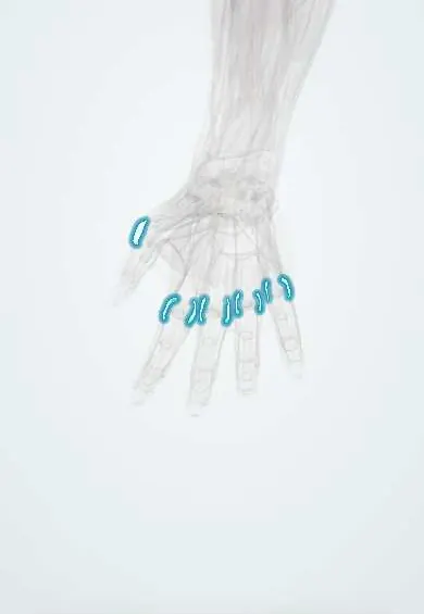
Joints and ligaments · Upper limb
Knuckle collateral ligaments !
The knuckle collateral ligaments sit on each side of the finger knuckles; they stop sideways bending and are tightest with the knuckles bent.
Official name: Collateral ligaments of the metacarpophalangeal (MCP) joints
Opens in the Durgical app, no sign-up needed.
What it is
Bands on each side of every knuckle joint.
What it does
Stop the fingers bending sideways; they are tightest when the knuckles are bent.
Key facts
- Attaches
- Dorsal tubercles of the metacarpal heads to the bases of the proximal phalanges
- Function
- Side-to-side stability of the knuckles
- Tight in
- MCP flexion (cam effect): splint knuckles bent
Relations
- Superficial
- Sagittal bands of the extensor mechanism and the interosseous tendons; the lumbrical tendon passes on the radial side
- In front
- Volar plate, where the accessory part inserts, and the A1 pulley with the flexor tendons
- Behind
- Extensor tendon over the joint, within the extensor mechanism
- Deep
- Joint capsule and the sides of the metacarpal head
How to find it
The MCP joint lines lie just distal to the knuckles, which stand out when the fist is made. With the MCP bent, each collateral is felt as firm tissue at the side of the joint; point tenderness there after a sideways injury suggests a sprain of that ligament.
Examination
- Collateral stress test with the MCP at about 90° of flexion, compared with the other hand: laxity or a soft end point suggests a tear of the proper collateral ligament
On imaging
PA and oblique X-rays look for avulsion fragments at the base of the proximal phalanx or the metacarpal head; the Brewerton view (knuckles flexed, back of the fingers on the plate, angled beam) shows small metacarpal head fractures and collateral avulsions. Ultrasound or MRI shows the ligament itself.
Clinical notes
- Bony avulsions from the base of the proximal phalanx are the X-ray clue; displaced or large fragments involve the joint surface
- In MCP joint replacement for rheumatoid arthritis, the radial collateral is often reconstructed to help correct ulnar drift
Development
In children the collaterals anchor to the epiphysis of the proximal phalanx, so a sideways force more often breaks through the growth plate (Salter–Harris II) than tears the ligament; the classic case is the base of the little finger ('extra-octave' fracture).
Related conditions
Test yourself
Think of the answer, then tap the question to see it.
In what position are the knuckle collateral ligaments tight, and how does that guide splinting?
In MCP flexion (cam effect), so the knuckles are splinted bent to stop the collateral ligaments shortening.
What are the two parts of each knuckle collateral ligament?
A proper part to the base of the proximal phalanx and an accessory part that fans palmarly into the volar plate.
How are the knuckle collateral ligaments stress-tested?
With the MCP flexed to about 90°, compared with the other hand; laxity or a soft end point suggests a tear of the proper collateral.
Educational information, not medical advice. For any symptom, see a health professional.
Its names
- Official Latin (TA2)
- ligamenta metacarpophalangea collateralia TA2 1836
- Official Spanish (DeCS)
- Ligamentos colaterales metacarpofalángicos
- English (TA2)
- Collateral ligaments of the metacarpophalangeal (MCP) joints
Related
- Thumb UCL
- Volar plates (knuckles)
- 1st metacarpal
- 2nd metacarpal
- 3rd metacarpal
- 4th metacarpal
- 5th metacarpal
- Proximal phalanx, thumb
Sources: Terminologia Anatomica (TA2); Anatomy, Shoulder and Upper Limb, Metacarpophalangeal Joints; Anatomy, Shoulder and Upper Limb, Hand Metacarpal Phalangeal Joint; Anatomy of the Human Body (20th ed.): Surface Anatomy of the Upper Extremity (Lea & Febiger (Gray's Anatomy, 1918), 1918) · adapted, Public domain.
Spotted a mistake?We are always improving for the community. Tell us what is wrong and our team will review it.Report a mistake
Representative model, for educational purposes only.