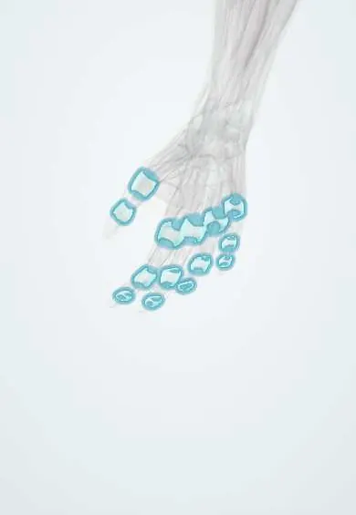
Rheumatoid arthritis of the hand and wrist !
Rheumatoid arthritis of the hand is an autoimmune joint inflammation of the wrists and knuckles on both sides; without treatment it deforms the hand.
Updated 2026-10-30
Structures involved
Tap one to see what it is and open it in 3D.
- Finger joint capsules
- Wrist capsule
- Knuckle collateral ligaments
- Extensor tendons
- Extensor tendon sheaths
- Flexor tendon sheaths
- Ulna
- Median nerve
- TFCC
In the clinical exam
Why it happens
- Chronic inflammation: causes, cells and features
- Innate and adaptive immunity: overview
- Synovial joints: capsule, synovial membrane and articular cartilage
What it is
Involvement of the hand and wrist by rheumatoid arthritis, a chronic autoimmune synovitis. It typically affects the wrists, the knuckle (MCP) joints and the middle finger (PIP) joints on both sides while sparing the end (DIP) joints, and untreated it leads to characteristic deformities.
Who gets it
Rheumatoid arthritis is more common in women and often starts between about 30 and 60 years of age; the hands and wrists are among the first and most often affected sites. It is a frequent reason for rheumatology care in Colombia and Latin America. Population surveys found a prevalence of 1.6% in Mexico and 0.94% in Luján, Argentina.
Causes
- Autoimmune inflammation of the synovium in a genetically predisposed person
- Environmental triggers, of which smoking is the best established
Risk factors
- Female sex
- Smoking
- Family history and genetic markers (shared epitope)
- Positive rheumatoid factor or anti-citrullinated protein antibodies, which also predict more erosive disease
Pathophysiology
Inflamed synovium (pannus) erodes cartilage and bone and stretches capsules, collateral ligaments and tendon sheaths. Tendons then slip and pull joints out of line: ulnar drift, swan neck, boutonnière. Synovitis over the distal ulna wears through tendons, and in the carpal tunnel it can compress the median nerve.
Classification
- Ulnar deviation and palmar subluxation of the fingers at the MCP joints
- Swan-neck deformity: PIP hyperextension with DIP flexion
- Boutonnière deformity: PIP flexion with DIP hyperextension, from attenuation of the central slip
- Thumb deformities (Z-thumb) and radial deviation of the wrist
- Caput ulnae: dorsal prominence of the ulnar head with extensor tendon rupture (Vaughan-Jackson lesion)
Symptoms and signs
- Symmetric pain and swelling of the wrists, MCP and PIP joints, with DIP joints usually spared
- Morning stiffness lasting more than about an hour
- Weak grip and difficulty with fine tasks
- Progressive deformities: ulnar drift, swan neck, boutonnière
- Sudden loss of finger extension from tendon rupture
- Rheumatoid nodules, numbness from carpal tunnel syndrome and fatigue
Diagnosis
- Clinical
- Symmetric polyarthritis of small hand joints with morning stiffness; the 2010 ACR/EULAR classification criteria combine joint count, serology, acute-phase reactants and symptom duration.
- Tests
- Rheumatoid factor and anti-citrullinated protein antibodies; ESR and CRP; blood count and liver and kidney tests before and during treatment.
- Imaging
- X-rays show periarticular osteopenia, marginal erosions and symmetric joint-space narrowing; ultrasound and MRI detect synovitis and erosions earlier.
Differential diagnosis
- Hand osteoarthritis (affects DIP joints, with Heberden nodes)
- Psoriatic arthritis
- Systemic lupus erythematosus arthropathy
- Gout or calcium pyrophosphate disease
- Viral arthritis, for example chikungunya
Goal of treatment
Achieve remission or low disease activity early, preserve joints and tendons, prevent deformity and keep hand function and independence.
Treatment
- Medical
- Disease-modifying antirheumatic drugs started early with a treat-to-target strategy: conventional synthetic agents first, adding biologic or targeted synthetic agents if the response is inadequate.
- Medical
- Short courses of glucocorticoids as a bridge, nonsteroidal anti-inflammatory drugs for symptoms, and intra-articular corticosteroid injection for single active joints.
- Rehabilitation
- Hand therapy: joint protection, splints for wrist and finger alignment, exercises and adaptive devices.
- Surgical
- Synovectomy and tenosynovectomy, tendon repair or transfer after rupture, distal ulna resection, MCP joint replacement and wrist fusion or replacement for advanced disease.
Complications
- Fixed deformities and loss of hand function
- Extensor or flexor tendon ruptures
- Carpal tunnel syndrome and trigger finger
- Joint destruction and wrist collapse
- Infections and other adverse effects of immunosuppression
Prognosis
With early disease-modifying treatment many patients reach remission or low activity and avoid deformity. Late or inadequate treatment leads to progressive joint and tendon damage; seropositive disease tends to be more erosive.
Prevention
- Stopping smoking
- Early referral of persistent symmetric small-joint arthritis for prompt treatment
Test yourself
Think of the answer, then tap the question to see it.
Which finger joints are typically spared in rheumatoid arthritis of the hand?
The distal interphalangeal (DIP) joints; rheumatoid arthritis mainly affects the wrists, MCP and PIP joints.
What is a swan-neck deformity?
Hyperextension of the PIP joint with flexion of the DIP joint.
What causes a boutonnière deformity in rheumatoid arthritis?
PIP synovitis stretches the central slip of the extensor mechanism, so the PIP flexes and the DIP hyperextends.
Which antibodies are most specific for rheumatoid arthritis?
Anti-citrullinated protein antibodies (anti-CCP).
What is the Vaughan-Jackson lesion?
Rupture of the extensor tendons of the ulnar-side fingers worn through over an eroded, dorsally prominent ulnar head.
Related conditions
- Rheumatoid arthritis
- Carpal tunnel syndrome
- Trigger finger
- Flexor tendon injury
- Systemic lupus erythematosus
- Gout
Educational information, not medical advice. For any symptom, see a health professional.
Sources: Hand and Wrist Rheumatoid Arthritis (StatPearls (NCBI Bookshelf), 2023); Burden of illness of rheumatoid arthritis in Latin America: a regional perspective (Clinical rheumatology, 2015) · adapted, CC BY 4.0.
Spotted a mistake?We are always improving for the community. Tell us what is wrong and our team will review it.Report a mistake
Representative model, for educational purposes only.