Joints and ligaments · Lower limb
Deep MCL !
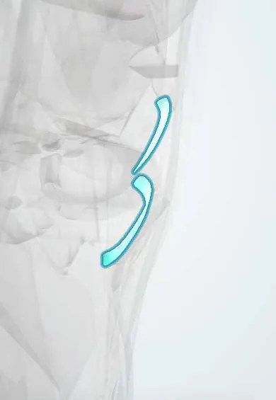
The deep MCL is the inner layer of the medial collateral ligament, joined to the knee capsule and the medial meniscus.
Official name: Deep part of tibial collateral ligament
Opens in the Durgical app, no sign-up needed.
What it is
The deeper layer of the MCL, joined to the joint capsule and the medial meniscus.
What it does
Keeps the knee from bending inward when it is pushed from the outside.
Key facts
- Attaches
- Thickened medial capsule: femur to medial meniscus (meniscofemoral part) and meniscus to tibia below joint line (meniscotibial part)
- Function
- Secondary restraint to valgus and external rotation; anchors medial meniscus, making it less mobile than the lateral
- Tight in
- Knee extension; external rotation of tibia
Relations
- Superficial
- Superficial MCL, separated from it by a small bursa
- Deep
- Peripheral rim of the medial meniscus, then the medial compartment of the joint
- Behind
- Posterior oblique ligament and the posteromedial capsule
How to find it
Not felt apart from the superficial MCL: both lie under the tender spot on the medial joint line. In a medial approach it appears once the superficial MCL is lifted; from inside the joint it is the capsular attachment of the meniscus, seen above and below its edge when the medial compartment is opened with valgus.
Examination
- Valgus stress at 30° (with the superficial MCL): increased medial opening; opening also at 0° means the posteromedial capsule is involved
On imaging
Coronal MRI: a thin dark band hugging the meniscus beneath the superficial MCL. A tear shows as fluid between meniscus and femur (meniscofemoral part) or meniscus and tibia (meniscotibial part); fluid between the meniscus and capsule suggests a meniscocapsular separation.
Common problems
- Sprain or tear from a blow to the outer side of the knee or a twisting fall. Graded from mild (1) to complete (3).
How it is diagnosed
An exam test that gently pushes the knee inward (valgus stress test). MRI if the injury is severe or others are suspected.
Treatment
- Most MCL injuries heal without surgery: rest, a hinged brace and physical therapy.
- Surgery is uncommon, mainly for severe or combined injuries.
Clinical notes
- Meniscocapsular separation: the meniscus pulls away from its capsular attachment in the well-vascularised periphery and can heal
- In knee replacement for a varus knee, the medial soft-tissue release usually begins with the deep MCL
- In arthroscopy, needle fenestration of the medial collateral complex (pie-crusting) can widen a tight medial compartment
Related conditions
Test yourself
Think of the answer, then tap the question to see it.
What are the two parts of the deep medial collateral ligament?
Meniscofemoral above the medial meniscus and meniscotibial (coronary) below it: a thickening of the medial capsule.
How does the deep MCL help explain why medial meniscus tears are more common than lateral ones?
It anchors the medial meniscus firmly, making it less mobile than the lateral meniscus.
Why can a meniscocapsular separation at the deep MCL heal?
The meniscus pulls away from its capsular attachment in the well-vascularised periphery.
Educational information, not medical advice. For any symptom, see a health professional.
Its names
- Official Latin (TA2)
- pars profunda ligamenti collateralis tibialis TA2 1898
- Official Spanish (DeCS)
- Porción profunda del ligamento colateral tibial DeCS D017888
- English (TA2)
- Deep part of tibial collateral ligament
- Also called
- deep part of medial collateral ligament of knee
Related
Sources: Terminologia Anatomica (TA2); DeCS; Anatomy, Bony Pelvis and Lower Limb, Knee Medial Collateral Ligament.
Spotted a mistake?We are always improving for the community. Tell us what is wrong and our team will review it.Report a mistake
Representative model, for educational purposes only.