Joints and ligaments · Lower limb
MCL !
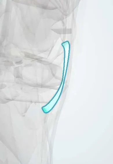
The medial collateral ligament (MCL) runs along the inner side of the knee and keeps it from bending inward.
Official name: Tibial collateral ligament
Opens in the Durgical app, no sign-up needed.
What it is
A broad band on the inner side of the knee, from the femur to the tibia.
What it does
Keeps the knee from bending inward when it is pushed from the outside.
Key facts
- Attaches
- Medial epicondyle of femur to medial condyle and upper medial shaft of tibia (tibial collateral ligament)
- Function
- Resists valgus stress; limits external rotation of tibia; most often injured knee ligament
- Tight in
- Knee extension; valgus stress
Relations
- Superficial
- Sartorius fascia; lower down the pes anserinus tendons (sartorius, gracilis, semitendinosus) cross it, with the pes anserine bursa between
- Deep
- Deep MCL (meniscofemoral and meniscotibial parts) and the medial meniscus, with a small bursa between the two layers
- Behind
- Posterior oblique ligament and the semimembranosus expansions, which together form the posteromedial corner
- Above
- Adductor tubercle with the adductor magnus tendon, and the femoral attachment of the MPFL just above and behind the medial epicondyle
How to find it
With the knee slightly bent, its line runs from the medial femoral epicondyle (the most prominent point on the inner side of the lower femur) across the joint line to the upper medial tibia; tenderness pinpoints whether the femoral end, the mid-portion or the tibial end is injured.
Examination
- Valgus stress at 30°: medial opening compared with the other knee; grade I about 0–5 mm, grade II 5–10 mm, grade III over 10 mm, usually without a firm end point
- Valgus stress at 0°: opening in full extension adds damage to the posteromedial capsule and often the ACL or PCL
On imaging
MRI grades the injury: fluid around an intact ligament (I), partial fibre disruption (II), complete discontinuity (III), and shows which end is torn. A distal tear can flip above the pes anserinus tendons (a Stener-like lesion) and heal poorly. Valgus stress X-rays compare medial opening with the other side; ultrasound shows the superficial layer well.
Common problems
- Sprain or tear from a blow to the outer side of the knee or a twisting fall. Graded from mild (1) to complete (3).
How it is diagnosed
An exam test that gently pushes the knee inward (valgus stress test). MRI if the injury is severe or others are suspected.
Treatment
- Most MCL injuries heal without surgery: rest, a hinged brace and physical therapy.
- Surgery is uncommon, mainly for severe or combined injuries.
Clinical notes
- Pellegrini–Stieda lesion: calcification near the femoral attachment on X-ray after an old MCL injury
- With combined MCL and ACL tears, the MCL is often allowed to heal first and the ACL is reconstructed afterwards
- Part of the unhappy triad (ACL, MCL, medial meniscus) after a blow to the outside of the knee with the foot planted
Related conditions
Test yourself
Think of the answer, then tap the question to see it.
At what knee position does valgus stress isolate the medial collateral ligament (MCL)?
At 30° of flexion. Opening also in full extension means the posteromedial capsule and often a cruciate are torn too.
What mechanism injures the MCL?
A blow to the outer side of the knee (valgus) or a twisting fall.
Why do most isolated MCL tears heal without surgery?
The MCL has a good blood supply, unlike the ACL.
What is the Pellegrini–Stieda lesion?
Calcification near the femoral attachment of the MCL on X-ray after an old MCL injury.
Which three structures make up the unhappy triad?
ACL, MCL and medial meniscus, after a blow to the outside of the knee with the foot planted.
Educational information, not medical advice. For any symptom, see a health professional.
Its names
- Official Latin (TA2)
- ligamentum collaterale tibiale; pars superficialis ligamenti collateralis tibialis TA2 1896
- Official Spanish (DeCS)
- Ligamento colateral medial de la rodilla DeCS D017888
- English (TA2)
- Tibial collateral ligament
- Also called
- medial collateral ligament of knee · superficial part of medial collateral ligament of knee · knee medial collateral ligament
Related
Sources: Terminologia Anatomica (TA2); DeCS; Anatomy, Bony Pelvis and Lower Limb, Knee Medial Collateral Ligament.
Spotted a mistake?We are always improving for the community. Tell us what is wrong and our team will review it.Report a mistake
Representative model, for educational purposes only.