Joints and ligaments · Lower limb
Medial meniscus !
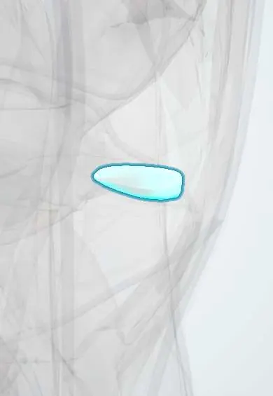
The medial meniscus is a C-shaped cartilage cushion on the inner side of the knee, between thigh and shin bones; it absorbs shock.
Opens in the Durgical app, no sign-up needed.
What it is
A C-shaped cushion of cartilage between the femur and tibia, on the inner side.
What it does
Spreads the body's weight across the joint, absorbs shock and helps keep the knee steady.
Key facts
- Where
- Medial tibial plateau, between medial femoral condyle and tibia; C-shaped, wider posteriorly
- Attaches
- Anterior and posterior horns to intercondylar area of tibia; firmly fixed to capsule and deep medial collateral ligament
- Function
- Deepens tibial surface; spreads load; absorbs shock; less mobile, so torn more often than lateral meniscus
Relations
- Above
- Medial femoral condyle
- Below
- Articular cartilage of the medial tibial plateau
- Medial
- Joint capsule through the coronary ligaments, and the deep layer of the medial collateral ligament
- Lateral
- Intercondylar area: the anterior horn attaches in front of the anterior cruciate ligament footprint and the posterior horn in front of the posterior cruciate ligament; the transverse ligament joins the two anterior horns
- Behind
- Semimembranosus sends fibres to the posterior capsule and posterior horn
How to find it
Not palpable itself. With the knee bent to about 90°, medial joint-line tenderness in the soft hollow between the medial femoral condyle and the tibial plateau points to it. At arthroscopy it is seen and probed through the anterior portals, with valgus stress opening the medial compartment.
Examination
- McMurray test: knee fully bent, tibia externally rotated with valgus stress, then extended; a painful click on the medial joint line suggests a medial meniscus tear
- Thessaly test: standing on the leg with the knee slightly bent and rotating the body; medial joint-line pain or catching is positive
- Apley grind test: prone, knee at 90°, pressing down while rotating causes pain; relief when the leg is pulled up instead points to the meniscus rather than a ligament
On imaging
MRI is the test of choice: a tear is high signal reaching an articular surface on proton-density images (on at least two slices), or a missing or displaced fragment (double PCL sign, flipped fragment). X-rays do not show the meniscus but are used for alignment and joint-space narrowing in degenerative tears.
Common problems
- Tears from twisting the bent knee, common in sports.
- Wear-related (degenerative) tears with age, sometimes with little injury.
- Symptoms: pain on the joint line, swelling, catching or locking.
How it is diagnosed
A physical exam (joint-line tenderness, special twisting tests such as McMurray). MRI shows the tear.
Treatment
- Without surgery: rest, physical therapy, anti-inflammatory medicine; injections in some wear-related cases.
- Surgery (usually arthroscopic): repairing the tear with stitches, or trimming the torn part (partial meniscectomy).
- Meniscal allograft transplant (a donor meniscus) for selected patients who have lost most of a meniscus.
Clinical notes
- Removing meniscus raises contact stress on the cartilage and speeds osteoarthritis, so repair is favoured for tears in the vascular zone
- Posterior root tears destroy hoop tension and behave like a total meniscectomy, with rapid cartilage loss
- Horizontal degenerative tears in middle-aged and older adults often accompany early osteoarthritis rather than cause the pain
Anatomical variants
- Discoid medial meniscus is rare, far rarer than the lateral form
Development
The menisci form within the developing knee before birth; in infants they are vascular throughout, and with age the blood supply withdraws to the outer rim.
Memory aid
Shapes: the medial meniscus is an open C, the lateral one almost an O.
Related conditions
Test yourself
Think of the answer, then tap the question to see it.
Which part of the medial meniscus is the commonest site of meniscal tears?
The posterior horn of the medial meniscus.
Why do tears in the inner part of the meniscus heal poorly?
Only the outer rim (red zone) is vascular; the inner white zone is avascular, which guides repair versus partial meniscectomy.
Why is the medial meniscus torn more often than the lateral meniscus?
It is firmly fixed to the capsule and the deep medial collateral ligament, so it is less mobile.
What is the classic McMurray test finding for a medial meniscus tear?
Knee fully bent, tibia externally rotated with valgus stress, then extended: a painful click on the medial joint line.
What does a bucket-handle tear of the medial meniscus cause, and what is its MRI sign?
The fragment flips into the intercondylar notch and locks the knee; on MRI it gives the double PCL sign.
Educational information, not medical advice. For any symptom, see a health professional.
Its names
- Official Latin (TA2)
- meniscus medialis TA2 1888
- Official Spanish (DeCS)
- Menisco medial DeCS D008592
- English (TA2)
- Medial meniscus
- Also called
- meniscus tibialis
Related
- Deep MCL
- Lateral meniscus
- Transverse ligament
- Meniscus roots
- Tibial cartilage
- Genicular arteries
- Femur
- Tibia
Sources: Terminologia Anatomica (TA2); DeCS; Anatomy, Bony Pelvis and Lower Limb: Medial Meniscus.
Spotted a mistake?We are always improving for the community. Tell us what is wrong and our team will review it.Report a mistake
Representative model, for educational purposes only.