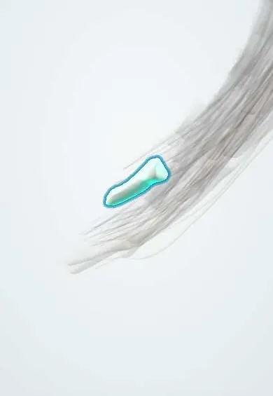
Distal phalanx, index finger !
The distal phalanx of the index finger is the bone of the fingertip; the deep flexor and the end of the extensor tendon attach to its base.
Official name: Distal phalanx of the index finger
Opens in the Durgical app, no sign-up needed.
What it is
The distal bone of the index finger. It carries the nail and the fingertip pad.
What it does
One of the finger bones; the joints between them let the finger curl.
Key facts
- Joints with
- Middle phalanx (DIP)
- Landmarks
- Base (FDP/FPL insertion, terminal extensor), tuberosity under the nail
Relations
- Above
- Head of the middle phalanx at the DIP joint
- In front
- FDP tendon to the base; the fingertip pulp, rich in digital nerve endings, used in fine pinch
- Behind
- Terminal extensor tendon at the base; the nail bed and germinal matrix
How to find it
The bone under the index fingertip and nail; the palmar pulp over it is the classic autonomous sensory area of the median nerve, used to check that nerve.
Examination
- Profundus test: with the middle phalanx held still, the person bends the fingertip; no active DIP flexion means the FDP tendon is cut or avulsed
On imaging
A lateral view shows a mallet fragment from the dorsal base and whether the distal phalanx has slipped palmarward; a tuft fracture is seen best on the PA view.
Clinical notes
- Mallet finger: the terminal extensor tears or avulses a dorsal fragment, leaving the DIP flexed
- Tuft fractures from crush injuries often come with nail bed injury
- Paronychia or a pulp abscess over it can reach the bone and cause osteomyelitis of the tuft when drainage is late
Development
Distal phalanges are among the first bones of the hand to ossify; the primary centre appears at the tip (the tuft) and a secondary centre forms at the base.
Related conditions
- Psoriasis
- Viral warts (verruca)
- Psoriatic arthritis
- Hypertrophic osteoarthropathy and digital clubbing
- Hand infections (paronychia, felon, flexor tenosynovitis)
Test yourself
Think of the answer, then tap the question to see it.
Which tendons insert on the distal phalanx of the index finger?
Flexor digitorum profundus on the palmar base and the terminal extensor tendon on the dorsal base.
Why can the index finger flex its DIP joint independently of the other fingers?
The index has its own flexor digitorum profundus muscle belly.
What is mallet finger?
The terminal extensor tears or avulses a dorsal fragment of the distal phalanx, leaving the DIP flexed.
Educational information, not medical advice. For any symptom, see a health professional.
Its names
- Official Latin (TA2)
- phalanx distalis manus TA2 1279
- Official Spanish (DeCS)
- Falange distal del índice
- English (TA2)
- Distal phalanx of the index finger
Related
Sources: Terminologia Anatomica (TA2); Anatomy, Shoulder and Upper Limb, Hand Bones; 8.2 Bones of the Upper Limb (OpenStax (Rice University), 2022) · adapted, CC BY 4.0; 8.5 Development of the Appendicular Skeleton (OpenStax (Rice University), 2022) · adapted, CC BY 4.0; Anatomy of the Human Body (20th ed.): Articulations of the Digits (Lea & Febiger (Gray's Anatomy, 1918), 1918) · adapted, Public domain.
Spotted a mistake?We are always improving for the community. Tell us what is wrong and our team will review it.Report a mistake
Representative model, for educational purposes only.