Psoriatic arthritis !
An inflammatory arthritis linked to psoriasis that can swell whole fingers, damage end joints and affect the spine and tendons.
Updated 2026-11-01
Structures involved
Tap one to see what it is and open it in 3D.
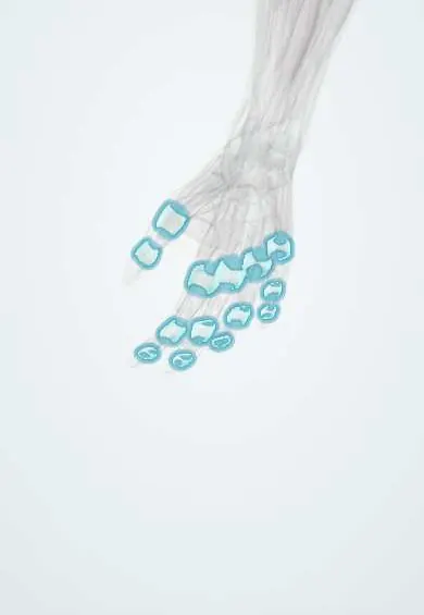
- Finger joint capsules
- Distal phalanx, index finger
- Sacroiliac ligaments
- Distal phalanx, middle finger
- Skin
- Achilles tendon
- Toe joint capsules
- Plantar fascia
In the clinical exam
Why it happens
- Chronic inflammation: causes, cells and features
- Bone remodeling
- Synovial joints: capsule, synovial membrane and articular cartilage
What it is
A chronic inflammatory arthritis of the spondyloarthritis family that occurs in people with psoriasis or a family history of it. It can involve peripheral joints, the spine, entheses, digits (dactylitis) and nails.
Who gets it
Develops in roughly one in five people with psoriasis, usually aged 30 to 50 and equally in men and women. In most cases skin psoriasis appears years before the arthritis. It is seen in Colombia and the rest of Latin America, where it is often underdiagnosed among dermatology patients.
Causes
- Immune-mediated; cause unknown
- Genetic predisposition (HLA-B27 in axial disease, HLA-C*06 with psoriasis)
- Possible triggers: joint trauma (deep Koebner phenomenon), infections, obesity
Risk factors
- Psoriasis, especially severe, scalp, intergluteal or nail psoriasis
- Family history of psoriasis or psoriatic arthritis
- Obesity
- Smoking
Pathophysiology
The IL-23/IL-17 axis and TNF drive inflammation that starts at entheses (tendon and ligament insertions) and spreads to synovium and bone. The disease both erodes bone and forms new bone, giving the typical pencil-in-cup deformity of distal joints, periostitis and spinal syndesmophytes. Nail disease reflects inflammation at the nail–enthesis complex of the distal phalanx.
Classification
- Asymmetric oligoarthritis (4 or fewer joints)
- Symmetric polyarthritis (rheumatoid-like)
- Distal interphalangeal-predominant arthritis
- Axial spondyloarthritis (sacroiliitis, spondylitis)
- Arthritis mutilans (severe destructive form)
Symptoms and signs
- Pain, swelling and morning stiffness of joints, often asymmetric
- Involvement of the distal finger joints next to nail changes
- Whole swollen sausage-like fingers or toes (dactylitis)
- Heel pain from Achilles or plantar fascia enthesitis
- Inflammatory back pain that improves with exercise
- Nail pitting, onycholysis and skin plaques of psoriasis
- Fatigue; red, painful eye (uveitis)
Diagnosis
- Clinical
- Inflammatory arthritis, enthesitis or spinal disease plus psoriasis (current, past or family), nail dystrophy, dactylitis and negative rheumatoid factor, as weighted in the CASPAR classification criteria.
- Tests
- No specific test. ESR and CRP may be normal or high; rheumatoid factor and anti-CCP are usually negative; HLA-B27 in axial disease. Uric acid may be raised, so joint fluid analysis helps exclude gout. Screening for tuberculosis and hepatitis before biologics.
- Imaging
- Radiographs show erosions with new bone formation, pencil-in-cup deformity, periostitis and asymmetric sacroiliitis or bulky syndesmophytes. Ultrasound and MRI detect early synovitis, enthesitis and sacroiliitis.
Differential diagnosis
- Rheumatoid arthritis
- Erosive hand osteoarthritis (Heberden nodes)
- Gout
- Reactive arthritis
- Ankylosing spondylitis
- Enteropathic arthritis
Goal of treatment
Reach remission or low disease activity across all domains (joints, spine, entheses, dactylitis, skin and nails), prevent joint damage and manage cardiovascular and metabolic comorbidity.
Treatment
- Medical
- Non-steroidal anti-inflammatory drugs and intra-articular glucocorticoids for symptom relief; systemic glucocorticoids are used sparingly because stopping them can flare psoriasis.
- Medical
- Conventional synthetic DMARDs (methotrexate, leflunomide, sulfasalazine) for peripheral arthritis; they do not help axial disease.
- Medical
- Biologic DMARDs (TNF, IL-17, IL-12/23 and IL-23 inhibitors), JAK inhibitors and PDE4 inhibitors for active or axial disease, enthesitis or poor response.
- Rehabilitation
- Physiotherapy, exercise and joint protection.
- Lifestyle
- Weight loss, stopping smoking and control of cardiovascular risk factors.
- Follow-up
- Monitoring of disease activity, liver tests and blood counts on methotrexate, and of infections on biologics after tuberculosis screening.
Drugs for this condition
Complications
- Permanent joint destruction and deformity (arthritis mutilans)
- Spinal fusion and reduced mobility
- Uveitis
- Cardiovascular disease, metabolic syndrome, fatty liver and diabetes
- Infections on immunosuppressive therapy
- Depression and reduced quality of life
Prognosis
Course ranges from mild to destructive. Early diagnosis and treat-to-target therapy limit damage; polyarticular onset, high inflammation and early erosions predict a worse outcome. Cardiovascular risk is increased.
Test yourself
Think of the answer, then tap the question to see it.
What is the classic radiographic deformity of psoriatic arthritis?
Pencil-in-cup deformity of the distal interphalangeal joints, with erosions plus new bone formation.
Which clinical features suggest psoriatic rather than rheumatoid arthritis?
DIP involvement, dactylitis, nail pitting, asymmetric pattern, enthesitis, axial disease and negative rheumatoid factor.
Why are systemic glucocorticoids used sparingly in psoriatic disease?
Their withdrawal can trigger a severe psoriasis flare, including pustular psoriasis.
What screening is required before starting a TNF inhibitor?
Screening for latent tuberculosis (and hepatitis B and C).
Related conditions
- Ankylosing spondylitis
- Reactive arthritis
- Rheumatoid arthritis
- Rheumatoid arthritis of the hand and wrist
- Gout
- Uveitis
- Crohn disease
- Ulcerative colitis
- Myocardial Infarction (Heart Attack)
Educational information, not medical advice. For any symptom, see a health professional.
Sources: Psoriatic arthritis (MedlinePlus (U.S. National Library of Medicine), 2025).
Spotted a mistake?We are always improving for the community. Tell us what is wrong and our team will review it.Report a mistake
Representative model, for educational purposes only.