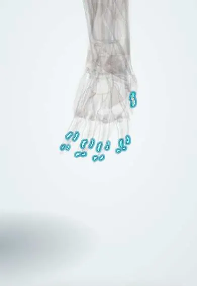
Joints and ligaments · Upper limb
Finger collateral ligaments !
The finger collateral ligaments sit on each side of the middle and end finger joints; they keep each joint working as a straight hinge.
Official name: Collateral ligaments of the finger joints
Opens in the Durgical app, no sign-up needed.
What it is
Small bands on each side of the finger joints; a sideways bend sprains or tears them (a 'jammed' finger).
What it does
Keep the finger joints working as straight hinges.
Key facts
- Attaches
- Heads of the proximal/middle phalanges to the bases of the next phalanges
- Function
- Side-to-side stability of PIP and DIP joints
- Tight in
- Throughout (accessory parts in extension)
Relations
- In front
- Volar plate, which receives the accessory fibres; flexor tendons in their sheath, with the A3 pulley at the PIP level
- Behind
- Lateral bands and central slip of the extensor mechanism
- Superficial
- Transverse retinacular and Cleland ligaments, then the digital nerves and digital arteries running along each side of the finger
How to find it
The IP joints are marked by the palmar finger creases and by the skin wrinkles on the back of the fingers. Each collateral is pressed on the side of the joint, in the middle of the side face; tenderness on one side with spindle-shaped swelling points to a sprain of that ligament.
Examination
- Collateral stress test of the PIP with the joint straight and at about 30° of flexion, compared with the neighbouring finger: wide opening (often quoted as more than 20°) suggests a complete tear
- Active range of motion after a digital block: a joint that stays congruent through active flexion and extension is considered stable
On imaging
A true lateral X-ray of the single finger is needed to see a subtle subluxation or fracture; PA and oblique views show avulsion fragments from the base of the middle phalanx, or condylar fractures that mimic a sprain.
Clinical notes
- Unicondylar fractures of the proximal phalanx head can look like a collateral sprain but are unstable
- Chronic laxity of the radial collateral of the index PIP weakens pinch, since it takes the thumb's pressure
- Scarring of the accessory collaterals and volar plate contributes to fixed PIP flexion contractures after injury
Test yourself
Think of the answer, then tap the question to see it.
Which finger joint is most often sprained, injuring its collateral ligaments?
The PIP joint; its collaterals have proper and accessory parts, like those of the knuckle.
What usually has to tear for gross instability of a finger joint?
The collateral ligament and at least part of the volar plate together.
Which fracture can mimic a finger collateral ligament sprain but is unstable?
A unicondylar fracture of the proximal phalanx head.
Educational information, not medical advice. For any symptom, see a health professional.
Its names
- Official Latin (TA2)
- ligamenta interphalangea collateralia manus TA2 1843
- Official Spanish (DeCS)
- Ligamentos colaterales interfalángicos de la mano
- English (TA2)
- Collateral ligaments of the finger joints
Related
- Volar plates (fingers)
- Distal phalanx, thumb
- Distal phalanx, index finger
- Distal phalanx, middle finger
- Distal phalanx, ring finger
- Distal phalanx, little finger
- Middle phalanx, index finger
- Middle phalanx, middle finger
Sources: Terminologia Anatomica (TA2); Anatomy, Shoulder and Upper Limb, Metacarpophalangeal Joints; Anatomy of the Human Body (20th ed.): Surface Anatomy of the Upper Extremity (Lea & Febiger (Gray's Anatomy, 1918), 1918) · adapted, Public domain.
Spotted a mistake?We are always improving for the community. Tell us what is wrong and our team will review it.Report a mistake
Representative model, for educational purposes only.