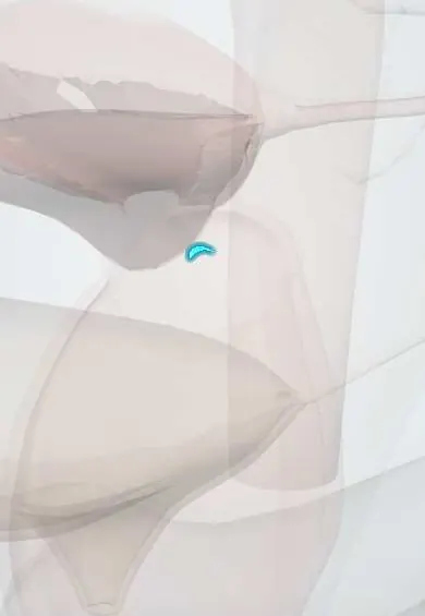
Reproductive system · Pelvis · Female
External os !
The external os is the small opening of the cervix into the vagina; cervical screening samples are taken around it, where most cervical cancers start.
Official name: External os of uterus
Opens in the Durgical app, no sign-up needed.
What it is
The opening of the cervix into the vagina, surrounded by the transformation zone where the inner and outer cell types meet.
What it does
Screening samples are taken here; most cervical cancers start in the transformation zone.
Key facts
- Where
- Opening of the cervical canal on the portio; round in nulliparous, slit-like after vaginal birth
- Function
- Surrounded by the transformation zone (squamocolumnar junction), site of HPV-related precancer; sampled in screening
Relations
- Above
- Cervical canal (endocervix), lined by columnar mucus-secreting epithelium, leading up to the internal os
- Lateral
- Ectocervix of the vaginal part, covered by stratified squamous epithelium, and beyond it the vaginal fornix
- Below
- Lumen of the vagina, facing its posterior wall
How to find it
Seen through a speculum as the central opening on the vaginal part of the cervix. For cytology a brush or spatula is rotated around it to sample the whole transformation zone; samples for HPV testing are also taken from the cervix (some programmes accept self-collected vaginal samples).
Examination
- Visual inspection with acetic acid (VIA): after dilute acetic acid is applied, abnormal epithelium turns white (acetowhite); used where cytology is hard to access
- Schiller test (Lugol iodine, VILI): healthy glycogen-rich squamous epithelium stains mahogany brown; areas that stay pale are columnar or abnormal
- Colposcopy: a magnified view of the transformation zone after acetic acid and iodine, to guide biopsy
On imaging
On sagittal transvaginal ultrasound the cervical canal is a thin bright line ending at the external os; in pregnancy cervical length is measured along the canal from the internal to the external os. Nabothian cysts appear as small fluid cysts near it. Sagittal T2 MRI stages cervical cancer, which usually starts at the transformation zone. Its surface is examined by colposcopy, not radiology.
Common problems
- Persistent HPV infection can cause precancerous changes and, over years, cervical cancer.
- Cervical insufficiency (opening too early in pregnancy).
How it is diagnosed
Screening: an HPV test and/or a Pap smear (cytology); colposcopy and biopsy if abnormal.
Treatment
- HPV vaccination prevents most cervical cancers.
- Precancer is removed with a small procedure (LEEP/cone).
Clinical notes
- Precancer is often treated by excising the transformation zone with a wire loop (LEEP/LLETZ) or by ablation
- Deep or repeated excisions shorten the cervix and are linked to a higher risk of preterm birth in later pregnancies
- After menopause the junction often lies inside the canal: colposcopy may not see the whole zone, and sampling has to reach into the canal
- Cervical stenosis after excision or radiotherapy can trap menstrual blood in the uterus (haematometra)
- In Colombia screening combines cytology, HPV testing and, in hard-to-reach areas, visual inspection with acetic acid and iodine
Anatomical variants
- Shape changes with childbirth: small and round in a woman who has not given birth vaginally, a transverse slit with an anterior and a posterior lip after a vaginal birth
Related conditions
Test yourself
Think of the answer, then tap the question to see it.
What is the transformation zone at the external os, and why does it matter?
The band of metaplastic squamous epithelium between the original and current squamocolumnar junction; HPV acts on its immature cells, so precancer (CIN) starts here.
In visual inspection with acetic acid (VIA), what indicates abnormal epithelium?
It turns white (acetowhite) after dilute acetic acid is applied.
In the Schiller test with Lugol iodine, which areas stay pale?
Columnar or abnormal epithelium; healthy glycogen-rich squamous epithelium stains mahogany brown.
Why can colposcopy be harder after menopause?
The squamocolumnar junction often lies inside the canal, so the whole transformation zone may not be seen and sampling must reach into the canal.
Educational information, not medical advice. For any symptom, see a health professional.
Its names
- Official Latin (TA2)
- ostium externum uteri TA2 3512
- Official Spanish (DeCS)
- Orificio externo del útero
- English (TA2)
- External os of uterus
- Also called
- orificium externum uteri · orificium externum canalis cervicis · ostium uteri
Related
Sources: Terminologia Anatomica (TA2); Anatomy, Abdomen and Pelvis: Female Internal Genitals; Anatomy, Abdomen and Pelvis: Uterus.
Spotted a mistake?We are always improving for the community. Tell us what is wrong and our team will review it.Report a mistake
Representative model, for educational purposes only.