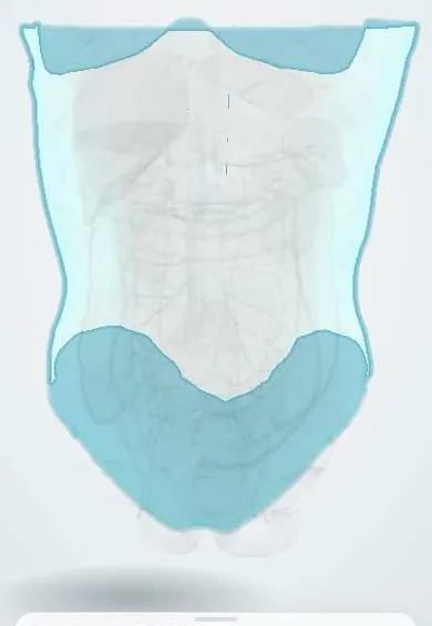
Superficial abdominal fascia !
These are the layers of connective tissue under the skin of the belly; below the navel they form a fatty layer and a firmer membrane that holds stitches.
Official name: Investing abdominal fascia
Opens in the Durgical app, no sign-up needed.
What it is
The thin fascia covering the outer abdominal muscles, under the fatty layer of the skin.
What it does
Wraps the muscles; it is closed in layers after surgery.
Key facts
- Where
- Subcutaneous: fatty superficial layer (Camper) over membranous deep layer (Scarpa); thin investing fascia directly on external oblique
- Attaches
- Scarpa fuses with fascia lata just below inguinal ligament; continues into perineum as Colles fascia and over penis/scrotum (dartos)
- Function
- Covers and wraps the wall muscles; Scarpa is sutured as a layer; it guides extravasated urine to abdominal wall and perineum, not the thigh
Relations
- Superficial
- Skin; the fatty Camper layer carries the superficial epigastric, superficial circumflex iliac and superficial external pudendal vessels, and the superficial inguinal lymph nodes in the groin
- Deep
- The thin investing fascia lies directly on the external oblique muscle and aponeurosis; at the superficial inguinal ring it continues onto the cord as the external spermatic fascia
- Above
- Above the umbilicus it becomes a single layer, continuous with the superficial fascia of the chest
- Below
- Scarpa fascia fuses with the fascia lata about a finger's breadth below the inguinal ligament; medially it passes into the perineum as Colles fascia and around the penis and scrotum, where the fat is replaced by the dartos muscle
How to find it
In a lower abdominal incision, Scarpa fascia appears as a whitish membrane inside the subcutaneous fat, under the yellow Camper fat and above the shiny external oblique aponeurosis.
On imaging
Not seen on its own on radiographs. On ultrasound and CT it is a thin bright line on the external oblique that separates subcutaneous fat from muscle. Its main use is to place a lesion: a hematoma, abscess or lipoma above it lies in the subcutaneous tissue, one below it lies in or under the muscle. In necrotizing infection, CT shows gas and fluid tracking along the fascial planes.
Common problems
- Hernias: a weak spot lets fat or bowel bulge through (groin, belly button, old incisions).
- Muscle strains.
How it is diagnosed
Examination, often while coughing or standing; ultrasound or CT if unclear.
Treatment
- Hernias that cause symptoms are repaired, usually with a mesh.
Clinical notes
- Straddle injury tearing the bulbar urethra: urine and blood fill the superficial perineal space, giving a butterfly-shaped perineal bruise and swelling of the scrotum, penis and lower wall, sparing the thighs
- Fournier gangrene spreads from the perineum along Colles fascia, dartos and Scarpa fascia; the extent seen on the skin often underestimates it
- Scarpa fascia is often closed as its own layer in lower abdominal incisions
- An ectopic testis may lie in the superficial inguinal pouch, between Scarpa fascia and the external oblique aponeurosis
Test yourself
Think of the answer, then tap the question to see it.
What are the two layers of the superficial abdominal fascia below the umbilicus?
The fatty Camper fascia superficially and the membranous Scarpa fascia deeply; Scarpa fascia holds sutures and is often closed as its own layer.
After a straddle injury tearing the bulbar urethra, why does urine spread up the abdominal wall but not into the thigh?
Scarpa fascia continues as Colles fascia and dartos but fuses with the fascia lata just below the inguinal ligament, closing the way to the thigh.
Along which planes can Fournier gangrene spread from the perineum?
Along Colles fascia, dartos and Scarpa fascia onto the abdominal wall; the extent seen on the skin often underestimates it.
Educational information, not medical advice. For any symptom, see a health professional.
Its names
- Official Spanish (DeCS)
- Fascia de revestimiento del abdomen DeCS D005205
- English (TA2)
- Investing abdominal fascia
Sources: DeCS; Anatomy, Abdomen and Pelvis: Abdominal Wall; Anatomy, Abdomen and Pelvis: Anterolateral Abdominal Wall Fascia.
Spotted a mistake?We are always improving for the community. Tell us what is wrong and our team will review it.Report a mistake
Representative model, for educational purposes only.