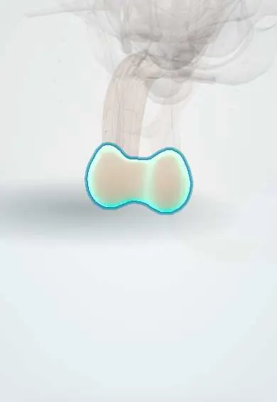
Reproductive system · Pelvis · Male
Scrotum !
The scrotum is the pouch of skin and muscle that holds the testes outside the body; it draws them up in the cold and relaxes in the heat to keep them cool.
Opens in the Durgical app, no sign-up needed.
What it is
A pouch of thin, wrinkled skin with a layer of smooth muscle (dartos), divided inside into two compartments, one for each testis.
What it does
Holds the testes outside the body and helps keep them cooler: the dartos and the cremaster draw them up in the cold and relax in the heat. Its lymph drains to the groin nodes, while the testes drain to nodes beside the aorta.
Key facts
- Where
- Skin pouch below the root of the penis, divided by a septum (raphe on the surface) into two compartments, each with a testis, epididymis and lower spermatic cord
- Attaches
- Layers: skin, dartos (continuous with Colles and Scarpa fascia), external spermatic fascia, cremaster, internal spermatic fascia, tunica vaginalis
- Function
- Keeps the testes about 2–3 °C below core temperature (dartos, cremaster, pampiniform plexus); skin lymph to superficial inguinal nodes, unlike the testis
Relations
- Above
- Root of the penis in front and the superficial inguinal rings, through which the spermatic cords enter
- Behind
- Perineum, with the bulb of the corpus spongiosum under the bulbospongiosus and the perineal body
- Lateral
- Medial surfaces of the thighs
- Contents
- On each side a testis, epididymis and the lower spermatic cord, separated by the scrotal septum
How to find it
Examined standing and lying in good light; each side is checked for contents, and any swelling is assessed for whether the examiner can get above it, whether light passes through it and whether it is separate from the testis.
Examination
- Getting above the swelling: if the upper edge of a scrotal swelling cannot be felt, it is probably an inguinoscrotal hernia
- Transillumination: hydroceles and spermatoceles glow; tumours, blood and most hernias do not
On imaging
Color Doppler ultrasound is the study of choice for the scrotum and its contents. The wall thickens in edema and infection. Classic findings: hydrocele (anechoic fluid around the testis), torsion (absent flow), epididymitis or orchitis (increased flow) and a testicular tumor (solid mass within the testis). CT shows gas in Fournier gangrene; MRI solves unclear cases.
Common problems
- Swellings: hydrocele, varicocele, hernia; any new lump should be checked.
- Skin infections and conditions.
How it is diagnosed
Examination; scrotal ultrasound.
Clinical notes
- A scrotal approach is used for vasectomy and hydrocele repair; a groin approach for a suspected testicular tumour
- Scrotal skin cancer in chimney sweeps was the first occupational cancer described (soot)
Anatomical variants
- Bifid scrotum (with severe hypospadias) and ectopic scrotum are rare
Development
Forms from the labioscrotal swellings, which fuse in the midline under androgens; the raphe marks the fusion. In the female the same swellings become the labia majora.
Related conditions
Test yourself
Think of the answer, then tap the question to see it.
Why does urine or blood from an anterior urethral injury fill the scrotum?
The scrotal dartos is continuous with Colles fascia.
What is Fournier gangrene, and why are the testes usually spared?
A fast necrotising infection of the perineum and scrotum along fascial planes, a surgical emergency; the testes have their own blood supply.
Which nerves supply the front and back of the scrotum?
Front: ilioinguinal and genital branch of genitofemoral (L1). Back: scrotal branches of the pudendal and posterior femoral cutaneous nerves (S2–S3).
What does it mean if the examiner cannot get above a scrotal swelling?
It is probably an inguinoscrotal hernia.
Educational information, not medical advice. For any symptom, see a health professional.
Its names
- Official Latin (TA2)
- scrotum
- Official Spanish (DeCS)
- Escroto
- English (TA2)
- Scrotum
Related
Sources: Anatomy, Abdomen and Pelvis, Scrotum.
Spotted a mistake?We are always improving for the community. Tell us what is wrong and our team will review it.Report a mistake
Representative model, for educational purposes only.