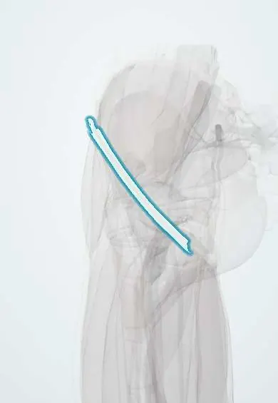
Inguinal ligament !
The inguinal ligament is a band along the groin crease; the main nerves and vessels of the thigh pass under it.
Opens in the Durgical app, no sign-up needed.
What it is
A band running along the groin crease, from the bony point at the front of the hip bone to the pubic bone.
What it does
Forms the floor of the inguinal canal. The femoral nerve, artery and vein pass under it into the thigh.
Key facts
- Attaches
- Anterior superior iliac spine to pubic tubercle; rolled lower border of external oblique aponeurosis
- Function
- Floor of inguinal canal; femoral vessels, femoral nerve and iliopsoas pass beneath it into thigh
Relations
- Course
- From the ASIS to the pubic tubercle, curving down toward the thigh; medially it turns back as the lacunar ligament and continues along the pecten as the pectineal (Cooper) ligament
- Above
- Inguinal canal, its gutter carrying the spermatic cord or round ligament; the deep ring above its midpoint
- Below
- Laterally the muscular space (iliopsoas, femoral nerve, lateral femoral cutaneous nerve); medially the femoral sheath with the femoral artery, vein and canal
- Medial
- Lacunar ligament, the medial border of the femoral ring
How to find it
Along a line from the ASIS to the pubic tubercle, slightly above the groin crease; the pubic tubercle is found by following the adductor longus tendon up to the pubis.
Examination
- Deep ring occlusion test: pressure over the deep ring while the person coughs; a hernia held back suggests indirect, one still bulging suggests direct (limited accuracy)
On imaging
Ultrasound is the main test for a groin lump: the ligament is followed from the ASIS to the pubic tubercle. A hernia above it, with Valsalva, is inguinal (lateral to the inferior epigastric vessels if indirect, medial if direct); below it and medial to the femoral vein, femoral. CT shows the same in complex cases. Radiographs only show its bony ends.
Common problems
- Inguinal and femoral hernias occur next to it.
Clinical notes
- Femoral artery puncture is made below the ligament, over the femoral head; a puncture above it risks retroperitoneal bleeding
- The lateral femoral cutaneous nerve passes under or through it near the ASIS; compression there causes meralgia paresthetica
- Open mesh hernia repair sutures the lower edge of the mesh to the shelving edge of the ligament
Development
It is the rolled-under lower edge of the external oblique aponeurosis. The inguinal canal above it forms when the gubernaculum leads the testis (or, in females, the round ligament) through the abdominal wall behind a pouch of peritoneum, the processus vaginalis; a processus that stays open underlies the indirect inguinal hernia.
Memory aid
NAVEL from lateral to medial below the ligament: Nerve, Artery, Vein, Empty space, Lymphatics (femoral canal).
Related conditions
Test yourself
Think of the answer, then tap the question to see it.
How are inguinal and femoral hernias distinguished in relation to the pubic tubercle?
Inguinal hernias appear above and medial to the pubic tubercle; femoral hernias below and lateral to it.
What is the order of structures beneath the inguinal ligament, from lateral to medial?
NAVEL: Nerve, Artery, Vein, Empty space, Lymphatics (femoral canal).
Why is femoral artery puncture made below the inguinal ligament?
A puncture above the inguinal ligament risks retroperitoneal bleeding; the safe site is below it, over the femoral head.
Which nerve passes under or through the inguinal ligament near the ASIS and causes meralgia paresthetica when compressed?
The lateral femoral cutaneous nerve.
Educational information, not medical advice. For any symptom, see a health professional.
Its names
- Official Latin (TA2)
- ligamentum inguinale TA2 2365
- Official Spanish (DeCS)
- Ligamento inguinal
- English (TA2)
- Inguinal ligament
- Also called
- Poupart's ligament · lateral ligament of Vesalius · Vesalius' ligament · fallopian ligament
Related
- Femoral artery
- Femoral vein
- Femoral nerve
- Anterior cutaneous nerves of the thigh
- Iliopsoas fascia
- Hip bone
Sources: Terminologia Anatomica (TA2); Anatomy, Abdomen and Pelvis: Inguinal Ligament (Crural Ligament. Poupart Ligament).
Spotted a mistake?We are always improving for the community. Tell us what is wrong and our team will review it.Report a mistake
Representative model, for educational purposes only.