Nervous system · Head and neck
Geniculate bodies !
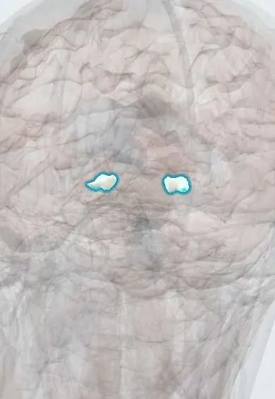
The geniculate bodies are two small bumps on the back of the thalamus; the lateral one relays vision and the medial one relays hearing to the cortex.
Official name: Lateral geniculate body
Opens in the Durgical app, no sign-up needed.
What it is
Two small bumps on the back of the thalamus.
What it does
Relay vision (lateral) and hearing (medial) to the cortex.
Key facts
- Where
- Under the back of the thalamus (pulvinar)
- Blood supply
- Posterior cerebral artery (thalamogeniculate branches), anterior choroidal artery
- Function
- Lateral: vision relay to the visual cortex (optic radiation); medial: hearing relay to the auditory cortex
Relations
- Above
- Pulvinar of the thalamus, which overhangs both bodies
- Medial
- The medial geniculate lies between the lateral one and the midbrain, joined to the inferior colliculus by its brachium; the superior colliculi lie further medially
- Course
- Vision: optic tract around the cerebral peduncle, lateral geniculate, optic radiation (lower fibres loop forward in the temporal lobe as Meyer's loop) to the visual cortex. Hearing: inferior colliculus, medial geniculate, acoustic radiation below the lentiform nucleus to the auditory cortex
How to find it
Two small swellings on the underside of the back of the thalamus, beneath the pulvinar: the lateral one where the optic tract ends, the medial one joined to the inferior colliculus by its brachium. They are seen on the side of the brainstem once the temporal lobe is lifted away.
Examination
- Confrontation visual fields: a homonymous field defect on the opposite side with brisk pupil reactions places the lesion at or behind the lateral geniculate
- Formal perimetry: a wedge-shaped homonymous defect suggests an infarct of the lateral geniculate
On imaging
Too small to judge on CT. High-resolution MRI shows them as two small bumps under the back of the thalamus (pulvinar): the lateral one at the end of the optic tract, the medial one nearer the midbrain. They are usually studied because of a field defect, such as a homonymous hemianopia; MRI then looks for an infarct, tumour or demyelination at this level.
Clinical notes
- Temporal lobe surgery or lesions cut Meyer's loop just beyond the lateral geniculate, giving an upper ('pie in the sky') quadrantanopia
- The lateral geniculate takes blood from both the anterior choroidal and the posterior choroidal arteries, which is why its infarcts can be patchy
Memory aid
Lateral = Light, Medial = Music.
Related conditions
Test yourself
Think of the answer, then tap the question to see it.
What do the lateral and medial geniculate bodies relay?
Lateral: vision to the visual cortex through the optic radiation; medial: hearing from the inferior colliculus to the auditory cortex.
Why are pupils normal with a lesion of the lateral geniculate body?
Pupillary light reflex fibres leave the optic tract for the pretectal area before the lateral geniculate.
Why can a lateral geniculate infarct give a wedge-shaped (sectoral) field defect?
It has a double blood supply, from the anterior and the posterior choroidal arteries, so its infarcts can be patchy.
Educational information, not medical advice. For any symptom, see a health professional.
Its names
- Official Latin (TA2)
- corpus geniculatum laterale TA2 5666
- Official Spanish (DeCS)
- Cuerpos Geniculados DeCS D005829
- English (TA2)
- Lateral geniculate body
Sources: Terminologia Anatomica (TA2); DeCS; Neuroanatomy, Nucleus Lateral Geniculate; Neuroanatomy, Thalamic Nuclei.
Spotted a mistake?We are always improving for the community. Tell us what is wrong and our team will review it.Report a mistake
Representative model, for educational purposes only.