Nervous system · Head and neck
Temporal lobe !
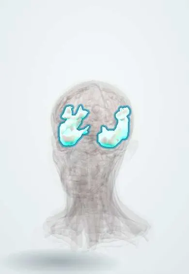
The temporal lobe is the lower side part of each half of the brain, behind the temple; it handles hearing, understanding language, memory and recognising faces.
Official name: Middle temporal gyrus
Opens in the Durgical app, no sign-up needed.
What it is
The lower side part of each hemisphere, behind the temple.
What it does
Hearing, language understanding, memory and recognising faces and objects.
Key facts
- Where
- Below the lateral sulcus
- Blood supply
- MCA (lateral), PCA (inferior)
- Function
- Hearing, language comprehension (Wernicke), memory, face recognition
Relations
- Above
- Frontal and parietal lobes across the lateral sulcus, in which the middle cerebral artery runs; the insula is hidden deep in the fissure
- Below
- Floor of the middle cranial fossa in front; the tentorium and the cerebellum behind
- Medial
- Uncus and parahippocampal gyrus beside the midbrain, with the oculomotor nerve and the posterior cerebral artery at the tentorial notch
- Lateral
- Squamous part of the temporal bone and the temporalis muscle
- Deep
- Temporal horn of the lateral ventricles, with the hippocampus in its floor and the amygdala at its tip
How to find it
On the lateral surface three parallel gyri (superior, middle and inferior) run back below the lateral sulcus; on the upper surface, hidden in the fissure, lie the transverse gyri of the auditory cortex. The inferior surface carries the fusiform and parahippocampal gyri. MRI for epilepsy uses thin coronal slices tilted perpendicular to the long axis of the hippocampus.
Examination
- Three-word recall after a few minutes of other tasks: words repeated correctly at first but lost later point to medial temporal (hippocampal) dysfunction
- Confrontation visual fields: a matching defect in the upper quadrants on the side opposite the lesion points to the Meyer loop
- Naming common objects: difficulty finding names (anomia) is frequent with lesions of the dominant temporal lobe
On imaging
Herpes simplex encephalitis shows asymmetric high T2/FLAIR signal and restricted diffusion in the medial temporal lobes, insula and orbital frontal cortex, typically sparing the basal ganglia. Mesial temporal sclerosis shows a small, bright hippocampus on coronal FLAIR.
Common problems
- Head injury and tumours here change personality, judgement and planning.
- Front-lobe dementia changes behaviour first.
How it is diagnosed
Neurological and memory tests; CT or MRI.
Treatment
- Surgery for tumours when possible.
- Rehabilitation and support.
Clinical notes
- Herpes simplex encephalitis targets the medial and inferior temporal lobes: fever, confusion, behaviour change and seizures
- Anterior temporal lobectomy for drug-resistant temporal lobe epilepsy can cut Meyer loop and leave an upper quadrant field defect
- Lesions of the fusiform gyrus (inferior temporo-occipital, often bilateral or right-sided) cause prosopagnosia, the inability to recognise familiar faces
- In Colombia and the rest of Latin America, neurocysticercosis (larval cysts of the pork tapeworm) is a common cause of acquired seizures; a cyst or calcified scar in the cortex of any lobe can be the focus
- Middle ear and mastoid infection can spread through the thin tegmen tympani to cause a temporal lobe abscess (otogenic abscess), with headache, fever and sometimes aphasia
Development
As the hemisphere enlarges it curves back and then down and forward in a C; the temporal lobe forms the lower limb of that C and carries the temporal horn, the hippocampus and the tail of the caudate with it.
Memory aid
"Pie in the sky": temporal lobe (Meyer loop, upper quadrant); "pie on the floor": parietal lobe (lower quadrant).
Related conditions
- MELAS (mitochondrial encephalomyopathy, lactic acidosis and stroke-like episodes)
- Traumatic brain injury
- Epilepsy and seizures
- Alzheimer disease
- Glioblastoma
- Local anaesthetic systemic toxicity (LAST)
- Viral encephalitis (herpes simplex encephalitis)
- Brain abscess
- Status epilepticus
- Frontotemporal dementia
- Autoimmune encephalitis (including anti-NMDA receptor encephalitis)
- Febrile seizures
- Schizophrenia
- Autism spectrum disorder (ASD)
- Schizoaffective, schizophreniform, brief psychotic and delusional disorders
Test yourself
Think of the answer, then tap the question to see it.
What is the typical sequence of a temporal lobe seizure?
An aura (rising epigastric feeling, fear, a smell or déjà vu), then a blank stare with lip-smacking or hand automatisms; the most common focal seizures in adults.
Which visual field defect follows damage to Meyer loop in the temporal lobe?
A contralateral superior quadrantanopia («pie in the sky»), as the lower optic radiation fibres loop forward around the temporal horn.
What happens in uncal herniation of the temporal lobe?
A mass above pushes the uncus down at the tentorial notch onto the oculomotor nerve, with a dilated pupil on the same side.
Which infection targets the medial and inferior temporal lobes, and how does it present?
Herpes simplex encephalitis: fever, confusion, behaviour change and seizures; MRI shows asymmetric high T2/FLAIR signal sparing the basal ganglia.
What does bilateral medial temporal damage cause?
Dense anterograde amnesia, as in the historical patient H.M. after bilateral resection in 1953.
Educational information, not medical advice. For any symptom, see a health professional.
Its names
- Official Latin (TA2)
- gyrus temporalis medius TA2 5495
- Official Spanish (DeCS)
- Lóbulo Temporal DeCS D013702
- English (TA2)
- Middle temporal gyrus
Related
Sources: Terminologia Anatomica (TA2); DeCS; Neuroanatomy, Temporal Lobe; 16.2 The Mental Status Exam (OpenStax (Rice University), 2022) · adapted, CC BY 4.0; 13.2 The Central Nervous System (OpenStax (Rice University), 2022) · adapted, CC BY 4.0; Anatomía humana (4.ª ed.), tomo 1, cap. 27: Relaciones del cerebro.
Spotted a mistake?We are always improving for the community. Tell us what is wrong and our team will review it.Report a mistake
Representative model, for educational purposes only.