Nervous system · Head and neck
Thalamus !
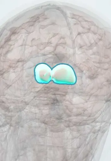
The thalamus is a pair of egg-shaped grey masses in the centre of the brain; it is the relay station through which almost all sensation reaches the cortex.
Opens in the Durgical app, no sign-up needed.
What it is
Two egg-shaped grey masses in the centre of the brain, beside the third ventricle.
What it does
The relay station: almost all sensation passes here on the way to the cortex.
Key facts
- Where
- Either side of the third ventricle
- Blood supply
- Posterior communicating (thalamotuberal branches: front); posterior cerebral (thalamoperforating: medial; thalamogeniculate: posterolateral, including VPL/VPM and geniculate bodies); posterior choroidal (upper surface, pulvinar); anterior choroidal (small share)
- Function
- Relay of all sensation except smell to the cortex; motor and arousal loops
Relations
- Medial
- Third ventricle, whose lateral wall it forms above the hypothalamic sulcus; in most brains the two thalami touch across it at the interthalamic adhesion
- Lateral
- Posterior limb of the internal capsule, and beyond it the lentiform nucleus (globus pallidus and putamen)
- Above
- Floor of the body of the lateral ventricle laterally; medially the fornix and the tela choroidea with the internal cerebral veins; the stria terminalis and thalamostriate vein mark its border with the caudate nucleus
- Below
- Hypothalamus in front, separated by the hypothalamic sulcus; subthalamus and midbrain tegmentum behind
- In front
- Anterior tubercle forming the back edge of the interventricular foramen
- Behind
- Pulvinar, overhanging the geniculate bodies and the superior colliculi, with the pineal gland in the midline
How to find it
Two egg-shaped grey masses forming the lateral walls of the third ventricle above the hypothalamic sulcus, often joined across it by the interthalamic adhesion. On axial MRI they lie medial to the posterior limb of the internal capsule, with the pulvinar jutting back over the midbrain.
On imaging
MRI shows thalamic infarcts best on diffusion images. Occlusion of an artery of Percheron gives matching paramedian infarcts on both sides, sometimes with the top of the midbrain. Symmetric medial thalamic signal around the third ventricle, with the mammillary bodies, suggests Wernicke encephalopathy.
Clinical notes
- Hypertensive thalamic haemorrhage: opposite-side sensory loss and weakness; the eyes may deviate down and in, as if looking at the nose
- Artery of Percheron occlusion: sudden drowsiness or coma, vertical gaze palsy and memory loss from infarction of both medial thalami
- Thiamine deficiency (often with alcohol) injures the medial thalami and mammillary bodies: Wernicke encephalopathy, then Korsakoff amnesia
- Stimulation of the ventral intermediate nucleus reduces essential tremor and tremor-dominant Parkinson disease
Anatomical variants
- The interthalamic adhesion is absent in a minority of brains, more often in men
- Artery of Percheron: a single trunk from one posterior cerebral artery (P1 segment) supplies both paramedian thalami, so one blockage infarcts both sides
Development
Comes from the diencephalon, the rear part of the forebrain vesicle, which also gives the hypothalamus and the optic cup; this shared origin is why the retina sends its fibres to the thalamus.
Memory aid
VPL = Limbs (body), VPM = Mouth (face); lateral geniculate = Light, medial geniculate = Music.
Related conditions
- Wernicke encephalopathy (thiamine deficiency)
- Sandhoff disease
- GM2 activator deficiency (GM2 gangliosidosis, AB variant)
- Intracerebral haemorrhage
- Hepatic encephalopathy
- Toxoplasmosis
- Vascular dementia
- Coma and brain death
- Essential tremor
- Perinatal asphyxia and hypoxic-ischaemic encephalopathy
- Alcohol withdrawal and delirium tremens
- Delirium
- Insomnia disorder
Test yourself
Think of the answer, then tap the question to see it.
Which thalamic nuclei relay touch from the body and from the face?
VPL for body touch and pain; VPM for the face and taste.
Which geniculate body of the thalamus relays vision and which relays hearing?
Lateral geniculate: vision; medial geniculate: hearing.
What is thalamic pain (Dejerine–Roussy) syndrome?
After an infarct of VPL/VPM, numbness on the opposite side is followed weeks to months later by burning pain and pain from light touch.
What does occlusion of an artery of Percheron cause?
Infarction of both medial thalami from one trunk: sudden drowsiness or coma, vertical gaze palsy and memory loss.
Which deficiency injures the medial thalami and mammillary bodies?
Thiamine deficiency, often with alcohol: Wernicke encephalopathy, then Korsakoff amnesia.
Educational information, not medical advice. For any symptom, see a health professional.
Its names
- Official Latin (TA2)
- thalamus TA2 5678
- Official Spanish (DeCS)
- Tálamo DeCS D013788
- English (TA2)
- Thalamus
- Also called
- dorsal thalamus · thalamencephalon
Related
Sources: Terminologia Anatomica (TA2); DeCS; Neuroanatomy, Thalamus; 13.1 The Embryologic Perspective (OpenStax (Rice University), 2022) · adapted, CC BY 4.0; 13.2 The Central Nervous System (OpenStax (Rice University), 2022) · adapted, CC BY 4.0.
Spotted a mistake?We are always improving for the community. Tell us what is wrong and our team will review it.Report a mistake
Representative model, for educational purposes only.