Nervous system · Head and neck
Midbrain !
The midbrain is the top part of the brainstem; it controls eye movements and pupil, hearing and visual reflexes, and carries movement pathways down.
Opens in the Durgical app, no sign-up needed.
What it is
The top part of the brainstem.
What it does
Eye movements, pupil reflexes, hearing and visual reflexes; carries the motor pathways down.
Key facts
- Where
- Top of the brainstem, through the tentorial notch
- Blood supply
- Paramedian (interpeduncular) branches of the basilar tip and PCA; collicular (quadrigeminal) and posterior choroidal arteries; SCA; a little from the anterior choroidal
- Function
- Nuclei of III and IV; pupil and eye-movement reflexes; corticospinal tracts pass
Relations
- In front
- Cerebral peduncles framing the interpeduncular fossa, where the oculomotor nerves emerge and the basilar artery divides into the posterior cerebral arteries
- Behind
- Tectum (superior and inferior colliculi) in the quadrigeminal cistern, under the pineal gland; the brachium of the superior colliculus runs to the lateral geniculate body and that of the inferior colliculus to the medial geniculate body; the trochlear nerves emerge just below the inferior colliculi
- Lateral
- Posterior cerebral and superior cerebellar arteries winding round it, with the oculomotor nerve between them; the free edge of the tentorium cerebelli and the uncus of the temporal lobe
- Above
- Thalamus and hypothalamus of the diencephalon
- Below
- Pons
- Contents
- Cerebral aqueduct ringed by periaqueductal grey; red nucleus, substantia nigra, oculomotor and trochlear nuclei, medial longitudinal fasciculus; in each crus, corticospinal fibres in the middle three-fifths, with frontopontine and corticonuclear fibres medial and temporo-parieto-pontine fibres lateral
How to find it
A short segment between the diencephalon and the pons that passes through the tentorial notch. In front, the two cerebral peduncles diverge around the interpeduncular fossa, where the oculomotor nerves come out; behind are the four colliculi. On midsagittal MRI the tectum lies behind the aqueduct and the tegmentum in front of it.
Examination
- Pupillary light reflex: the efferent limb is the oculomotor nerve via the Edinger–Westphal nucleus; a unilateral fixed dilated pupil in a drowsy patient points to oculomotor compression
- Vertical gaze: loss of upward gaze with eyelid retraction suggests a lesion of the dorsal midbrain (Parinaud syndrome)
On imaging
Axial MRI shows the classic 'Mickey Mouse' outline, the peduncles as ears. In progressive supranuclear palsy the upper midbrain shrinks, giving a 'hummingbird' profile on sagittal MRI.
Clinical notes
- Downward shift of the brainstem in herniation tears small perforating arteries, causing Duret haemorrhages in the midbrain and pons
- Kernohan notch: the opposite peduncle is pressed against the tentorium, giving weakness on the same side as the mass, a false localising sign
- The periaqueductal grey modulates pain, a target of opioid effects
Development
Comes from the mesencephalon, the one primary vesicle that does not divide again; its narrow cavity becomes the cerebral aqueduct. Congenital aqueduct stenosis is a classic cause of hydrocephalus, with large lateral and third ventricles and a normal fourth.
Related conditions
- Pyruvate dehydrogenase deficiency
- Leigh syndrome
- Raised intracranial pressure and brain herniation
- Coma and brain death
- Brainstem stroke syndromes (Wallenberg and others)
- Progressive supranuclear palsy
- Traumatic brain injury
- Migraine
- Epidural haematoma
- Parkinson disease
Test yourself
Think of the answer, then tap the question to see it.
What is Weber syndrome?
A stroke in the base of the midbrain: oculomotor palsy on the same side (drooping lid, eye down and out, large pupil) with weakness of the opposite arm and leg.
What is unique about the trochlear nerve as it leaves the midbrain?
It is the only cranial nerve to leave the back of the brainstem, and it crosses first, so its nucleus serves the opposite superior oblique.
What does uncal herniation compress at the midbrain?
The oculomotor nerve (one fixed, dilated pupil) and the cerebral peduncle.
What is the Kernohan notch phenomenon?
The opposite cerebral peduncle is pressed against the tentorium, giving weakness on the same side as the mass, a false localising sign.
What does the midbrain look like on sagittal MRI in progressive supranuclear palsy?
The upper midbrain shrinks, giving a «hummingbird» profile.
Educational information, not medical advice. For any symptom, see a health professional.
Its names
- Official Latin (TA2)
- mesencephalon TA2 5874
- Official Spanish (DeCS)
- Mesencéfalo DeCS D008636
- English (TA2)
- Midbrain
Related
Sources: Terminologia Anatomica (TA2); DeCS; Neuroanatomy, Mesencephalon Midbrain; 13.1 The Embryologic Perspective (OpenStax (Rice University), 2022) · adapted, CC BY 4.0; 13.2 The Central Nervous System (OpenStax (Rice University), 2022) · adapted, CC BY 4.0; Tratado de anatomía humana (9.ª ed.), tomo II, Libro V, sección 2 (Encéfalo), capítulos I-V: bulbo, protuberancia, cerebelo, cuarto ventrículo, mesencéfalo.
Spotted a mistake?We are always improving for the community. Tell us what is wrong and our team will review it.Report a mistake
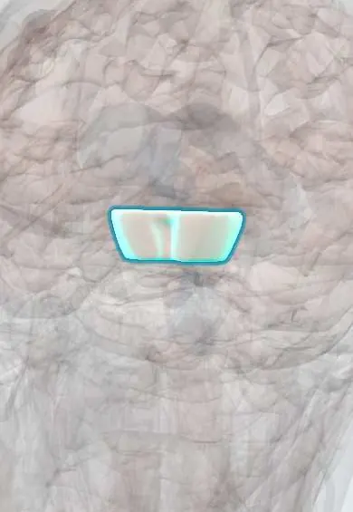
Representative model, for educational purposes only.