Nervous system · Head and neck
Visual cortex !
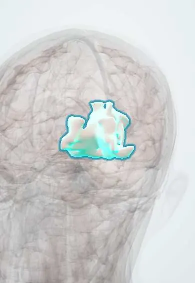
The visual cortex lies on the inner surface of the occipital lobe at the back of the brain; it is the first cortical stop for vision from both eyes.
Official name: Cuneus
Opens in the Durgical app, no sign-up needed.
What it is
The cortex around the calcarine sulcus on the inner surface of the occipital lobe.
What it does
First cortical stop for vision; a stroke here blanks half the visual field of both eyes.
Key facts
- Where
- Banks of the calcarine sulcus (occipital)
- Blood supply
- PCA (calcarine branch)
- Function
- Primary visual cortex (area 17); opposite visual field
Relations
- Course
- Input from the lateral geniculate body through the optic radiation: upper fibres through the parietal lobe, lower fibres around the temporal horn (Meyer loop)
- Above
- Cuneus, above the calcarine sulcus
- Below
- Lingual gyrus, below the calcarine sulcus
- In front
- Junction with the parieto-occipital sulcus; the splenium of the corpus callosum lies just in front
- Behind
- Occipital pole, where the macular area may reach onto the lateral surface
How to find it
It lines the calcarine sulcus on the medial surface of the occipital lobe, from the occipital pole forward to where the sulcus meets the parieto-occipital sulcus. Sagittal and coronal MRI show it best; most of it is buried in the walls of the sulcus.
Examination
- Confrontation fields and formal perimetry: a congruous homonymous defect with macular sparing suggests an occipital lesion
- Pupillary light reflex: normal in cortical blindness, because the reflex fibres leave the optic tract for the midbrain before the geniculate body
On imaging
Sagittal and coronal MRI show the calcarine sulcus and its two banks. A posterior cerebral artery infarct is bright on DWI in the medial occipital lobe and matches a homonymous hemianopia; a spared occipital pole can explain macular sparing. CT shows bleeds and older infarcts. Symmetric parieto-occipital oedema on FLAIR suggests posterior reversible encephalopathy syndrome (PRES).
Clinical notes
- Visual migraine aura, a shimmering zigzag that spreads over minutes, is attributed to a wave of spreading depression across the visual cortex
- Anton syndrome: bilateral damage with cortical blindness that the person denies, describing scenes that are not seen
Memory aid
The cuneus sits upstairs and sees the floor; the lingual gyrus sits downstairs and sees the sky.
Related conditions
Test yourself
Think of the answer, then tap the question to see it.
Where is the primary visual cortex?
Lining the calcarine sulcus on the medial surface of the occipital lobe (area 17), from the occipital pole forward.
Which bank of the calcarine sulcus receives the upper visual field?
The lower bank (lingual gyrus); the upper bank (cuneus) receives the lower field, so a cuneus lesion gives an inferior quadrantanopia.
Why is the visual cortex called striate cortex?
After the stria of Gennari, a band of myelinated fibres in layer IV visible to the naked eye.
Educational information, not medical advice. For any symptom, see a health professional.
Its names
- Official Latin (TA2)
- cuneus TA2 5485
- Official Spanish (DeCS)
- Corteza Visual Primaria DeCS D000087642
- English (TA2)
- Cuneus
- Also called
- striate cortex · V1
Related
Sources: Terminologia Anatomica (TA2); DeCS; Neuroanatomy, Visual Cortex.
Spotted a mistake?We are always improving for the community. Tell us what is wrong and our team will review it.Report a mistake
Representative model, for educational purposes only.