Cardiovascular system · Head and neck
Posterior cerebral artery !
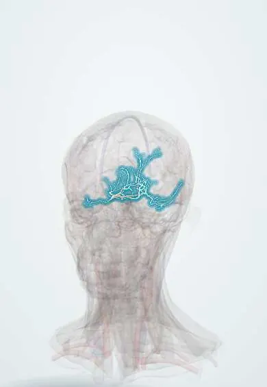
The posterior cerebral artery feeds the back of the brain, where vision is processed; a stroke here can cause loss of half the field of vision.
Opens in the Durgical app, no sign-up needed.
What it is
The end branches of the basilar artery, curving back around the midbrain.
What it does
Feeds the occipital lobe and lower temporal lobe; a stroke here causes loss of half the visual field.
Key facts
- Origin
- End of the basilar artery
- Branches
- Paramedian and thalamic perforators (interpeduncular); collicular (quadrigeminal) artery; posterior choroidal; temporal, calcarine, parieto-occipital
- Supplies
- Occipital lobe (visual cortex, cuneus), inferior temporal lobe except the pole, splenium of the corpus callosum; deep: posterior two thirds of the thalamus, geniculate bodies, midbrain (cerebral peduncle, red nucleus)
Relations
- Course
- From the basilar bifurcation in the interpeduncular cistern (P1) it receives the posterior communicating artery, winds back around the cerebral peduncle above the tentorium (P2) and reaches the medial occipital lobe, where it gives the calcarine and parieto-occipital branches
- Below
- Oculomotor nerve, which passes between it and the superior cerebellar artery; further back, the free edge of the tentorium cerebelli
- Medial
- Midbrain (cerebral peduncle), with the trochlear nerve running alongside in the ambient cistern
How to find it
It cannot be felt. It curves back around the midbrain, above the tentorial edge, alongside the oculomotor nerve, which leaves the brainstem between this artery and the superior cerebellar artery; its cortical branches run on the medial and lower surfaces of the occipital and temporal lobes.
Examination
- Confrontation visual fields: homonymous loss on the opposite side, often with central vision preserved
On imaging
Diffusion-weighted MRI shows the infarct in the occipital lobe, medial temporal lobe or thalamus; CT or MR angiography shows the occlusion.
Clinical notes
- Uncal herniation can pinch the artery against the tentorium and cause an occipital infarct
- Infarcts of the medial temporal lobe (hippocampus) can cause memory loss
Anatomical variants
- Fetal-type artery: it fills mainly from the internal carotid through a large posterior communicating artery, with a small P1
- Artery of Percheron: a single P1 branch feeds both paramedian thalami, so its blockage gives bilateral thalamic infarcts with sudden drowsiness or coma
Development
In the embryo it is a branch of the internal carotid; the basilar artery takes over later, and when the carotid origin persists it becomes the fetal-type artery.
Related conditions
Test yourself
Think of the answer, then tap the question to see it.
What visual deficit follows a posterior cerebral artery infarct of the occipital lobe, and why is the macula spared?
Opposite homonymous hemianopia; the occipital pole also receives blood from the middle cerebral artery.
What is alexia without agraphia, and which artery is involved?
The person can write but not read; a dominant posterior cerebral artery infarct involving the splenium.
What is Anton syndrome?
Cortical blindness denied by the person, after both posterior cerebral arteries are blocked, for example at the basilar tip.
What does occlusion of the artery of Percheron cause?
Bilateral paramedian thalamic infarcts with sudden drowsiness or coma; it is a single P1 branch of the posterior cerebral artery.
Educational information, not medical advice. For any symptom, see a health professional.
Its names
- Official Latin (TA2)
- arteria posterior cerebri TA2 4565
- Official Spanish (DeCS)
- Arteria Cerebral Posterior DeCS D020769
- English (TA2)
- Posterior cerebral artery
Related
Sources: Terminologia Anatomica (TA2); DeCS; Neuroanatomy, Posterior Cerebral Arteries.
Spotted a mistake?We are always improving for the community. Tell us what is wrong and our team will review it.Report a mistake
Representative model, for educational purposes only.